Françoise Cattin – författare
Visar alla böcker från författaren Françoise Cattin. Handla med fri frakt och snabb leverans.
10 produkter
10 produkter
Inbunden, Engelska, 2016
2 016 kr
Skickas inom 10-15 vardagar
This clinically oriented book will familiarize the reader with all aspects of the diagnosis of tumors and other disorders of the pituitary gland by means of magnetic resonance imaging (MRI). The coverage includes acromegaly, Cushing’s disease, Rathke cleft cysts, prolactinomas, incidentalomas, nonsecreting adenomas, other lesions of the sellar area, hypophysitis, and central diabetes insipidus. Normal radiologic anatomy and the numerous normal variants are described, and guidance is also provided on difficulties, artifacts, and other pitfalls. The book combines concise text and high-quality images with a question and answer format geared toward the needs of the practitioner. MRI is today considered the cornerstone in the diagnosis of diseases of the hypophyseal-hypothalamic region but the relatively small size of the pituitary gland, its deep location, the many normal anatomic variants, and the often tiny size of lesions can hinder precise evaluation of the anatomic structures and particularly the pituitary gland itself. Radiologists and endocrinologists will find MRI of the Pituitary Gland to be full of helpful information on this essential examination, and the book will also be of interest to internists and neurosurgeons.
E-bok
PDF, Engelska, 20161 737 kr
Läs direkt efter köp
This clinically oriented book will familiarize the reader with all aspects of the diagnosis of tumors and other disorders of the pituitary gland by means of magnetic resonance imaging (MRI). The coverage includes acromegaly, Cushing’s disease, Rathke cleft cysts, prolactinomas, incidentalomas, nonsecreting adenomas, other lesions of the sellar area, hypophysitis, and central diabetes insipidus. Normal radiologic anatomy and the numerous normal variants are described, and guidance is also provided on difficulties, artifacts, and other pitfalls. The book combines concise text and high-quality images with a question and answer format geared toward the needs of the practitioner. MRI is today considered the cornerstone in the diagnosis of diseases of the hypophyseal-hypothalamic region but the relatively small size of the pituitary gland, its deep location, the many normal anatomic variants, and the often tiny size of lesions can hinder precise evaluation of the anatomic structures and particularly the pituitary gland itself. Radiologists and endocrinologists will find MRI of the Pituitary Gland to be full of helpful information on this essential examination, and the book will also be of interest to internists and neurosurgeons.
Häftad, Engelska, 2018
1 458 kr
Skickas inom 10-15 vardagar
This clinically oriented book will familiarize the reader with all aspects of the diagnosis of tumors and other disorders of the pituitary gland by means of magnetic resonance imaging (MRI). The coverage includes acromegaly, Cushing’s disease, Rathke cleft cysts, prolactinomas, incidentalomas, nonsecreting adenomas, other lesions of the sellar area, hypophysitis, and central diabetes insipidus. Normal radiologic anatomy and the numerous normal variants are described, and guidance is also provided on difficulties, artifacts, and other pitfalls. The book combines concise text and high-quality images with a question and answer format geared toward the needs of the practitioner. MRI is today considered the cornerstone in the diagnosis of diseases of the hypophyseal-hypothalamic region but the relatively small size of the pituitary gland, its deep location, the many normal anatomic variants, and the often tiny size of lesions can hinder precise evaluation of the anatomic structures and particularly the pituitary gland itself. Radiologists and endocrinologists will find MRI of the Pituitary Gland to be full of helpful information on this essential examination, and the book will also be of interest to internists and neurosurgeons.
Häftad, Engelska, 1990
566 kr
Skickas inom 10-15 vardagar
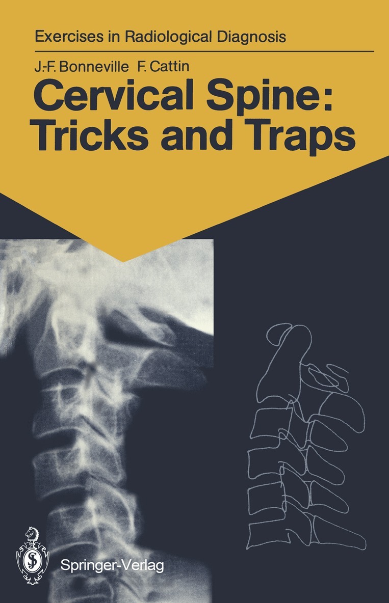
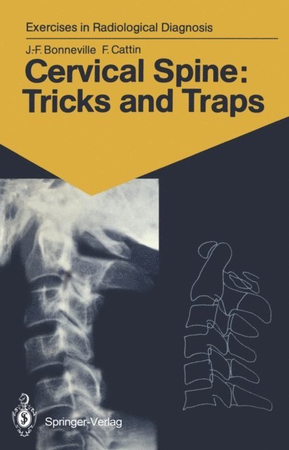
The cervical spine is always examined initially using standard radiographs, which often provide a sufficient basis for diagno- sis. Malformations, tumours, more frequently trauma, rheuma- tism, and even plain neck pain require a radiological exam- ination. The interpretation of radiological images is often difficult because of overlapping pieces of bone, the summation phe- nomena and the diversity of projections. In this book, two- or three-dimensional CTscans accompany the standard radiographs, serving as an excellent aid for com- prehension. It is almost as if the reader actually had the bones shown in the radiographs in his hands. From then on, everything becomes easy, superimpositions vanish, traps come to light, anatomy triumphs, and the images takes on life. Besanl$on, 1990 J .-F. BONNEVILLE, F. CATTIN v Contents Introduction 1 Iconography and Text with Corresponding Schemes 2 Subject Index ...123 VII Normal cervical spine: 3-D imaging 1 1 2 Case 1 1 Our starting point is a normal lateral radiograph of the cervical spine. It will serve as a guide and point of reference in our task of comprehending the interrelationships between the structures of the cervical spine.This radiograph correctly includes the entire area from the base of the skull to the cervicothora- cic junction. The soft tissues are clearly visible anteriorly, as are the extremities of the spinous processes posteriorly. In this case, with the subject looking straight ahead, is a slight, regular, physiological lordosis ofthe cervical column.
634 kr
Skickas inom 10-15 vardagar
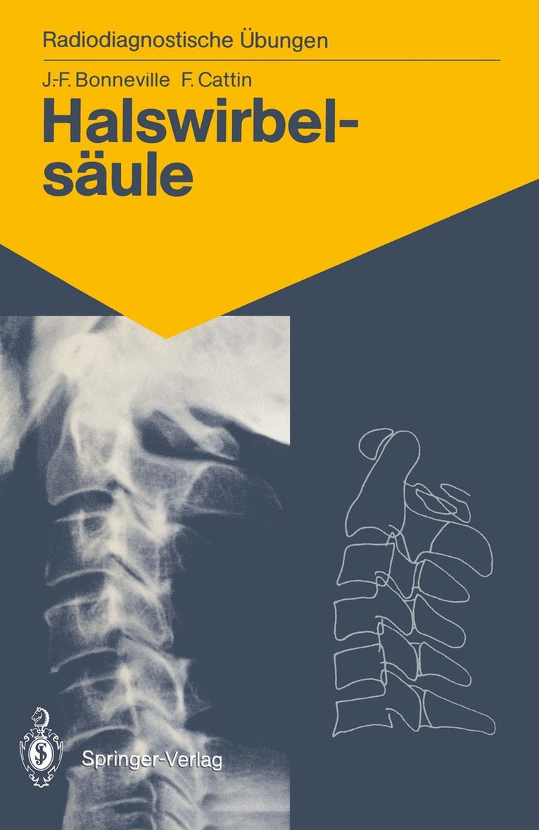
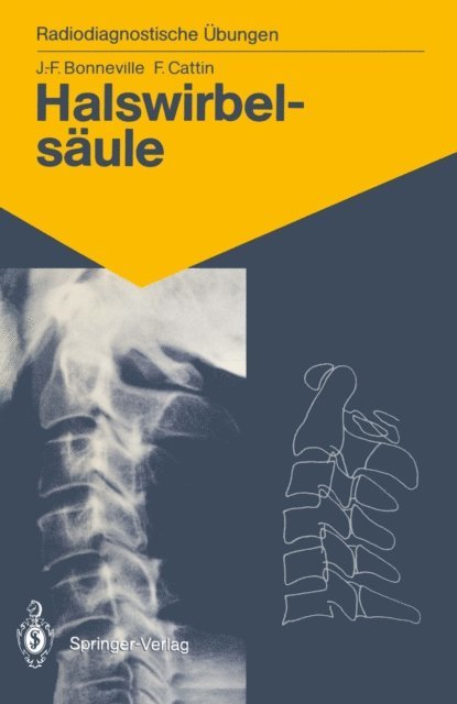
Erste Untersuchungen der Halswirbelsäule werden stets mit Hilfe von Röntgenaufnahmen durchgeführt, und in den meisten Fällen genügen diese als Grundlage für die Diagnose. Mißbildungen, Tumoren, und noch öfter Traumata, Rheuma und sogar ganz gewöhnliche Nackenschmerzen erfordern eine radiologische Untersuchung der Wirbelsäule. Die Auswertung jedoch ist schwierig. Nimmt man einen Halswirbel in die Hand, so stellt man fest, um welch komplexes Gebilde es sich hierbei handelt. Bei radiologischen Aufnahmen wird die Auswertung noch durch die sich überlappenden Knochenteile, Anhäufungen und die verschiedenen Blickwinkel erschwert. Das Buch von J.-F. Bonneville und F. Cattin stellt eine originelle Interpretationsmethode von Röntgenaufnahmen vor, die die Auswertung wesentlich erleichtert. Dieses Buch zeigt, daß zwei- bzw. dreidimensionale Computertomogramme eine ausgezeichnete Hilfe zum Verständnis von konventionellen Röntgenbildern sein können. Der Leser bekommt gleichsam Zugang zu jedem Einzelteil des Knochens, und von da an wird alles einfach, Überlappungen verschwinden, die in der Röntgenaufnahme verborgenen Tücken werden sichtbar, die Anatomie triumphiert, das Bild lebt. Die Halswirbelsäule von J.-F. Bonneville und F. Cattin ist unentbehrlich für jeden Radiologen in seiner täglichen Praxis, aber ebenso auch für Chirurgen, Rheumatologen und Physiotherapeuten, die sich für die Halswirbelsäule interessieren.
Inbunden, Engelska, 2013
1 899 kr
Skickas inom 5-8 vardagar
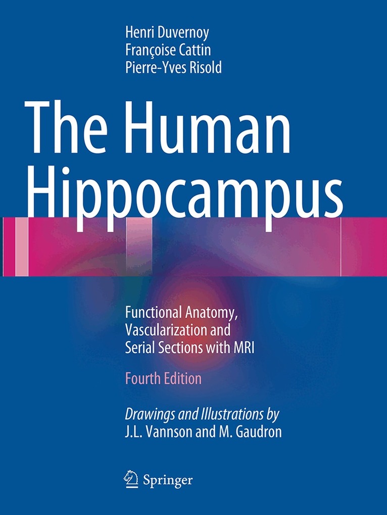
This new edition, like previous ones, offers a precise description of the anatomy of the human hippocampus based upon neurosurgical progress and the wealth of medical imaging methods available. The first part describes the fine structures of the hippocampus and is illustrated with new original figures. A survey is then provided of current concepts explaining the functions of the hippocampus, and the external and internal hippocampal vascularization is precisely described. The last and main part of the book presents serial sections in coronal, sagittal, and axial planes; each section is accompanied by a drawing to explain the MR 3T images. The new edition is also enriched by several MRI views of major hippocampal diseases. This comprehensive atlas of human hippocampal anatomy will be of interest to all neuroscientists, including neurosurgeons, neuroradiologists, and neurologists.
E-bok
PDF, Engelska, 20131 722 kr
Läs direkt efter köp
This new edition, like previous ones, offers a precise description of the anatomy of the human hippocampus based upon neurosurgical progress and the wealth of medical imaging methods available. The first part describes the fine structures of the hippocampus and is illustrated with new original figures. A survey is then provided of current concepts explaining the functions of the hippocampus, and the external and internal hippocampal vascularization is precisely described. The last and main part of the book presents serial sections in coronal, sagittal, and axial planes; each section is accompanied by a drawing to explain the MR 3T images. The new edition is also enriched by several MRI views of major hippocampal diseases. This comprehensive atlas of human hippocampal anatomy will be of interest to all neuroscientists, including neurosurgeons, neuroradiologists, and neurologists.
E-bok
PDF, Engelska, 2012734 kr
Läs direkt efter köp
E-bok
PDF, Tyska, 2013503 kr
Läs direkt efter köp
Erste Untersuchungen der Halswirbelsäule werden stets mit Hilfe von Röntgenaufnahmen durchgeführt, und in den meisten Fällen genügen diese als Grundlage für die Diagnose. Mißbildungen, Tumoren, und noch öfter Traumata, Rheuma und sogar ganz gewöhnliche Nackenschmerzen erfordern eine radiologische Untersuchung der Wirbelsäule. Die Auswertung jedoch ist schwierig. Nimmt man einen Halswirbel in die Hand, so stellt man fest, um welch komplexes Gebilde es sich hierbei handelt. Bei radiologischen Aufnahmen wird die Auswertung noch durch die sich überlappenden Knochenteile, Anhäufungen und die verschiedenen Blickwinkel erschwert. Das Buch von J.-F. Bonneville und F. Cattin stellt eine originelle Interpretationsmethode von Röntgenaufnahmen vor, die die Auswertung wesentlich erleichtert. Dieses Buch zeigt, daß zwei- bzw. dreidimensionale Computertomogramme eine ausgezeichnete Hilfe zum Verständnis von konventionellen Röntgenbildern sein können. Der Leser bekommt gleichsam Zugang zu jedem Einzelteil des Knochens, und von da an wird alles einfach, Überlappungen verschwinden, die in der Röntgenaufnahme verborgenen Tücken werden sichtbar, die Anatomie triumphiert, das Bild lebt. Die Halswirbelsäule von J.-F. Bonneville und F. Cattin ist unentbehrlich für jeden Radiologen in seiner täglichen Praxis, aber ebenso auch für Chirurgen, Rheumatologen und Physiotherapeuten, die sich für die Halswirbelsäule interessieren.
Häftad, Engelska, 2013
1 346 kr
Skickas inom 10-15 vardagar
This new edition, like previous ones, offers a precise description of the anatomy of the human hippocampus based upon neurosurgical progress and the wealth of medical imaging methods available. The first part describes the fine structures of the hippocampus and is illustrated with new original figures. A survey is then provided of current concepts explaining the functions of the hippocampus, and the external and internal hippocampal vascularization is precisely described. The last and main part of the book presents serial sections in coronal, sagittal, and axial planes; each section is accompanied by a drawing to explain the MR 3T images. The new edition is also enriched by several MRI views of major hippocampal diseases. This comprehensive atlas of human hippocampal anatomy will be of interest to all neuroscientists, including neurosurgeons, neuroradiologists, and neurologists.