Inbunden, Engelska, 2025
Diagnostic Ultrasound: Musculoskeletal
3707 kr
Beställningsvara. Skickas inom 11-20 vardagar. Fri frakt över 249 kr.
Beskrivning
**Selected for 2025 Doody’s Core Titles� in Radiologic Technology**
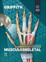
Develop a solid understanding of ultrasound and evolving musculoskeletal ultrasound practices with this multiple award-winning point-of-care reference in the popular Diagnostic Ultrasound series. Written by leading experts in the field, the third edition of Diagnostic Ultrasound: Musculoskeletal offers detailed, clinically oriented coverage of anatomy, techniques, and diagnoses in this complex area. Featuring more than 3,800 images and 200+ online videos, this edition showcases today’s rapidly evolving musculoskeletal ultrasound practice and its expanding applications for everyday clinical use. More than 100 detailed, clinically-oriented chapters provide expert guidance on ultrasound anatomy, technique, diagnosis, differential diagnosis, reporting, and ultrasound-guided interventional procedures for the entire musculoskeletal system.
Develop a solid understanding of ultrasound and evolving musculoskeletal ultrasound practices with this multiple award-winning point-of-care reference in the popular Diagnostic Ultrasound series. Written by leading experts in the field, the third edition of Diagnostic Ultrasound: Musculoskeletal offers detailed, clinically oriented coverage of anatomy, techniques, and diagnoses in this complex area. Featuring more than 3,800 images and 200+ online videos, this edition showcases today’s rapidly evolving musculoskeletal ultrasound practice and its expanding applications for everyday clinical use. More than 100 detailed, clinically-oriented chapters provide expert guidance on ultrasound anatomy, technique, diagnosis, differential diagnosis, reporting, and ultrasound-guided interventional procedures for the entire musculoskeletal system.
Produktinformation
- Utgivningsdatum: 2025-04-01
- Mått: 216 x 276 x 53 mm
- Vikt: 3 220 g
- Format: Inbunden
- Språk: Engelska
- Antal sidor: 1 088
- Upplaga: 3
- Förlag: Elsevier Health Sciences
- ISBN: 9780443285646
Utforska kategorier
Betyg & recensioner
0 recensioner
Inga recensioner tillgängliga.