• Fri frakt över 249 kr
  • •
  • Snabba leveranser
  • •
  • Billiga böcker
Kundservice

Du är på sajten för privatpersoner.

Företag, bibliotek eller offentlig verksamhet?

Du handlar på classic.bokus.com, där alla dina funktioner finns intakta.
Till classic.bokus.com
Bokus logotyp. Gå till startsidan.
  • Erbjudanden
  • Nyheter
  • Student
  • Topplistor
  • Barn & ungdom
  • Bokus Play
  • E-böcker
  • Pocketböcker
  • Spel & pussel

10% rabatt på allt med kod: NYSTART10 →

Sidfot

Mina sidor

    Hjälp

    • Kundservice
    • Vanliga frågor och svar
    • Frakt och leverans
    • Retur vid ångerrätt
    • Reklamera vara
    • Betalning
    • Köpvillkor
    • Allmänna villkor
    • Information om webbplatsens tillgänglighet

    Om Bokus

    • Om oss
    • Pressrum
    • För studenter
    • För företag
    • För bibliotek och offentlig verksamhet
    • För leverantörer
    • Hållbarhet

    Populärt

    • Aktuella erbjudanden
    • Presentkort
    • Studentlitteratur
    • Nya böcker
    • Topplistor
    • Signerade böcker
    • Engelska böcker

    Inspiration

    • Boktips
    • BookTok
    • Populära bokserier
    • Barnbokskaraktärer
    • Populära författare
    Logotyp för Bokus
    Följ oss på Facebook (extern länk)Följ oss på Instagram (extern länk)Följ oss på YouTube (extern länk)Följ oss på TikTok (extern länk)
    bokus @ CookiesAnpassa cookiesIntegritetspolicyKöpvillkor
    Till Citymail hemsida (extern länk)Till Budbee hemsida (extern länk)Till Postnord hemsida (extern länk)Till Schenker hemsida (extern länk)Till Early Bird hemsida (extern länk)Till Walleys hemsida (extern länk)
    1. Medicin
    2. Veterinärmedicin
    3. Veterinärmedicin: smådjur

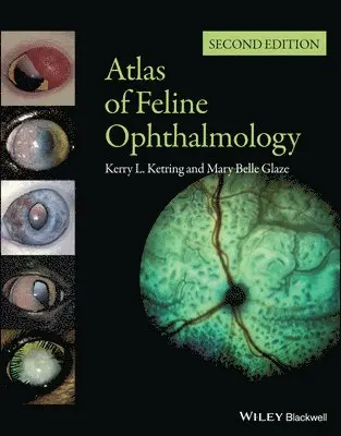
    Atlas of Feline Ophthalmology

    AvKerry L. Ketring,Mary Belle Glaze

    Inbunden, Engelska, 2012

    1 172 kr

    Beställningsvara. Skickas inom 5-8 vardagar. Fri frakt över 249 kr.

    Beskrivning

    Atlas of Feline Ophthalmology Successful management of eye disease relies on the veterinarian’s ability to identify ocular features and distinguish pathologic changes. Atlas of Feline Ophthalmology, Second Edition is an invaluable diagnostic reference, providing high-quality color photographs for comparison with a presenting complaint. Presenting 394 photographs illustrating both normal and pathologic ocular conditions, this Second Edition offers a current, complete reference on ocular diseases, adding conditions recognized since publication of the first edition, a broader geographic scope, and many new images with improved quality. Carefully designed for easy reference, the contents are divided into sections corresponding to specific anatomical structures of the eye. A useful appendix new to this edition groups figures by etiology, making it easy to find every image associated with a specific agent or disease. Atlas of Feline Ophthalmology, Second Edition is a useful tool aiding general practitioners in diagnosing eye disease in cats. Key Features Provides a compendium of high-quality color images of the feline eyeFocuses on the identification of a global list of diseases, conditions, and injuries affecting the eyeIncludes both normal and pathologic ocular conditionsActs as a diagnostic tool, with therapeutic information omitted by designAids clinicians in using ocular examinations to diagnose systemic diseaseDesigned for easy reference, with the book divided into sections according to ocular anatomical structures and an appendix grouping figures by etiology

    Produktinformation

    • Utgivningsdatum:2012-02-28
    • Mått:231 x 274 x 15 mm
    • Vikt:658 g
    • Format:Inbunden
    • Språk:Engelska
    • Antal sidor:192
    • Upplaga:2
    • Förlag:John Wiley and Sons Ltd
    • ISBN:9780470958742

    Utforska kategorier

    • Veterinärmedicin: smådjur inom Medicin

    Mer om författaren

    The Authors Kerry L. Ketring, DVM, DACVO, a veterinary ophthalmologist, was in private practice in Ohio and Kentucky for 32 years; he retired to Michigan, where he continues to see clients. He also lectures nationally and internationally. Mary Belle Glaze, DVM, MS, DACVO, is a veterinary ophthalmologist at the Gulf Coast Animal Eye Clinic, a private referral practice in Houston, Texas. Prior to that she was on staff at Louisiana State University’s Veterinary Teaching Hospital for 20 years and is a past president of the American College of Veterinary Ophthalmologists. She also lectures nationally and internationally.

    Recensioner i media

    "This is a superb update of the previous edition. The beautiful photographs will give veterinary practitioners new tools for identifying and treating feline ophthalmic disease. This book will be very useful for both general practitioners and veterinary ophthalmologists.”  (Doody’s, 30 August 2012)“In summary, the ‘Atlas of Feline Ophthalmology’ provides a comprehensive collection of pictures of feline ocular diseases and is a useful addition to the library of veterinary ophthalmologists and veterinarians with a deeper interest in ophthalmology.”  (Veterinary Record, 18 August 2012)

    Innehållsförteckning

    • Listing of Breed Predispositions to Ocular Disease . . . page xviiI. Normal EyeA. Diagrams1. Cross-sectional 12. Fundus oculus 2B. Normal adnexa/anterior segment1. Frontal view 3, 42. Lateral viewa. Lens and cornea 5b. Gross angle 63. Iridocorneal angle—Gonioscopic view 7C. Normal fundus 8–17II. Globe–Orbit RelationshipA. Convergent strabismus 18B. Enophthalmos1. Microphthalmia 192. Phthisis bulbi 203. Horner’s syndrome 214. Retrobulbar tumor 225. Pain 37, 40, 42, 76, 125, 143C. Exophthalmos1. Cellulitis/Retrobulbar abscess 23–252. Neoplasiaa. Retrobulbar lymphoma 26b. Zygomatic osteoma 273. Orbital pseudotumor 28, 29D. Proptosis 30E. Orbitalmucocele 31, 32III. AdnexaA. Eyelid agenesis 33–36B. Entropion 37C. Ectropion 38D. Distichiasis 39E. Blepharitis1. Herpetic 40, 542. Allergic blepharitis 41, 42, 513. Bacterial blepharitis 434. Meibomianitis 445. Demodicosis 496. Mycobacterial dermatitis 507. Food allergy 528. Pemphigus erythematosus 539. Persian idiopathic facial dermatitis 55F. Apocrine cystadenoma 45, 46G. Chalazion 47H. Lipogranulomatous conjunctivitis 48I. Granuloma/Histoplasmosis 68J. Neoplasia1. Cutaneous histiocytosis 562. Squamous cell carcinoma 57–593. Adenocarcinoma 60, 614. Mast cell tumor 62–645. Melanoma 656. Periorbital lymphoma 667. Nerve sheath tumor 67IV. ConjunctivaA. Dermoid 36, 69, 70B. Symblepharon72–75, 102C. Conjunctivitis1. Infectiousa. Herpesvirus 76, 77, 83b. Chlamydophila 78–80, 84c. Bartonella 81, 83, 84d. Mycoplasma 82e. Polymicrobial 83, 84f. Ophthalmia neonatorum 71g. Leishmania 88h. Blastomycosis 89i. Histoplasmosis 902. Allergica. Insect sting 85b. Drug reaction 423. Eosinophilic 86, 87, 104, 1514. Traumatic945. Conjunctival cysts 92, 936. Parasitic-Thelaziasis 95D. Dacryocystitis 96E. Neoplasia1. Lymphoma 912. Melanoma 97, 98V. Nictitating MembraneA. Nictitans protrusion1. Idiopathic prolapsed nictitating membrane 992. Glandular prolapse 1003. Everted cartilage 1014. Symblepharon 102, 1135. Horner’s syndrome 216. Abscess 1037. Retrobulbar neoplasia 228. Phthisis bulbi 209. Pain 37, 40, 42, 76, 125, 143B. Eosinophilic conjunctivitis 104C. Neoplasia1. Fibrosarcoma 1052. Squamous cell carcinoma 106, 1073. Lymphoma 1084. Plasmacytoma 109VI. CorneaA. Corneal opacities1. Persistent pupillary membranes 110–112, 171, 1722. Adherent leukoma 113, 158, 1593. Corneal degeneration 114, 1154. Florida spots 116, 1175. Storage disease (MPS-VI) 1186. Relapsing polychondritis 119B. Congenital Endothelial Dysfunction 123C. Keratoconus 120, 121D. Manx dystrophy 122E. Infectious keratitis1. Viral keratitis-Herpetica. Punctate 124b. Dendritic 125, 126c. Geographic 107, 127–131, 147, 1532. Mycoplasma 132, 1333. Bacteriala. Staphylococcus 134b. Pseudomonas 1354. Fungala. Candida 136b. Aspergillus 1375. Mycobacterial 138, 139F. Ulcerative keratitis1. Superficial ulcerationa. Keratoconjunctivitis sicca 25, 140, 153, 175, 176b. Neurotrophic 1402. Bullous keratitis 1413. Bullous keratopathy 1424. Descemetocele 143, 1445. Iris prolapse 35, 145G. Corneal Laceration 146H. Eosinophilic keratitis 107, 147–151I. Corneal sequestration 37, 152–155J. Foreign body 156K. Staphyloma 157–159L. Neoplasia1. Limbal melanocytoma (Scleral shelf melanoma, Epibulbar melanoma) 160–1622. Neuroblastic 1633. Squamous cell carcinoma 164VII. Anterior UveaA. Dyscorias1. Iris coloboma 36, 165, 1662. Corectopia 1673. Idiopathic dyscoria 1684. D-shaped pupil 1695. Spastic pupil syndrome 170B. Persistent pupillary membranes 110–112, 171, 172, 272C. Chediak-Higashi syndrome 173D. Iris atrophy 174E. Dysautonomia 175, 176F. Iris cysts/Iridocilary cysts 177–179, 267G. Anterior uveitis1. Iris abscess 1842. Virala. Feline leukemia complex/ Lymphoma 180–183, 185–190b. FIV 190, 191c. FIP 192–1963. Toxoplasmosis 197–2024. Fungala. Histoplasmosis 202–206b. Cryptococcosis 207, 208c. Blastomycosis 209, 210d. Coccidioidomycosis 2115. Bartonellosis 213–216, 2446. Polymicrobial 190, 202, 2127. Parasitica. Dirofilariasis 217b. Myiasis 2188. Metabolic/Hypertensiona. Hyperlipidemia 219b. Systemic hypertension 220, 2219. Trauma 222, 22310. Lens Induceda. Phacolytic 276b. Septic lens implantation 22411. Neoplasiaa. Feline diffuse iris melanoma (FDIM) 225, 226, 229, 231, 269, 270b. Iris melanoma 227, 228c. Iris amelanotic melanoma 227, 230, 232d. Iridociliary adenoma 233–236e. Spindle cell tumor 237f. Iridociliary leiomyoma 238g. Iridociliary leiomyosarcoma 239, 240h. Metastatic mammary adenocarcinoma 241i. Squamous cell carcinoma 242j. Metastatic hemangiosarcoma 243k. Primitive neural epithelial tumor 244l. Post-traumatic sarcoma 245, 24612. Post-inflammatory sequelaea. Lens capsule pigmentation 247, 250b. Posterior synechia/Iris bombé 248, 249c. Cataract 247, 248, 250d. Anterior lens luxation 286e. Iris cysts 179f. Glaucoma 264–266, 268–270VIII. GlaucomaA. Congenital/Goniodysgenesis 251, 252, 261, 262, 288, 388B. Inherited/Primary Open Angle Glaucoma (POAG)1. Siamese 2532. Domestic shorthair 254–257C. Feline Aqueous Humor Misdirection Syndrome (FAHMS) 258–260D. Secondary1. Post-inflammatory/Infectious 209, 224, 263–265, 2892. Systemic Hypertension 2663. Iridocilary cysts 2674. Neoplastica. Spindle cell tumor 237b. Lymphoma 190, 268c. Feline diffuse iris melanoma (FDIM) 232, 269, 270, 389IX. LensA. Senile nuclear sclerosis 271B. Cataract1. Congenital/Persistent pupillary membranes 272, 274–2772. Nutritional 2733. Inherited 173, 2754. Cataract resorption 277–279, 2815. Trauma/Post-inflammatory 245–248, 280–2826. Hypocalcemic 285C. Cataract classification by involvement1. Incipient 224, 247, 248, 258, 272, 273, 282, 2852. Immature 173, 239, 247, 248, 274, 275, 280, 2833. Mature 250, 2604. Hypermature/Phacolytic uveitis 276, 2845. Cataract resorption 19, 277–279, 281D. Encephalitozoon cuniculi 283, 284E. Lens luxation1. Anterior 259, 260, 286, 288, 2892. Posterior 254, 255, 257, 2873. Subluxation 160, 256X. VitreousA. Persistent hyaloid 288B. Vitreous hemorrhage 292C. Hyalitis1. FIV 2892. Toxoplasmosis 2903. Pyogranulomatous inflammation 2914. Retinal detachment/Systemic hypertension 292XI. Retina and choroidA. Congenital1. Cardiovascular Anomalies 2932. Coloboma 294, 2953. Retinal Folds 296, 297B. Chorioretinitis-Infectious1. Feline leukemia complex 298–3012. Panleukopenia 3023. Feline infectious peritonitis 303–3084. Fungal conditionsa. Histoplasmosis 309–313, 386b. Cryptococcosis 314–319c. Blastomycosis 320–322d. Coccidioidomycosis 323, 3245. Toxoplasmosis 325–3316. Feline hemotropic mycoplasmosis (feline infectious anemia) 3327. Bacterial 3338. Ophthalmomyiasis 334, 335C. Chorioretinitis-Traumatic 336, 349, 350D. Hypertensive retinopathy 337–347E. Retinal detachment1. Renal failure/Systemic hypertension 338, 342–3452. Trauma 3483. Neoplasia 373, 374, 3764. Infectious 308, 312, 313, 318, 321, 323, 331F. Retinal folds1. Dysplastic 296, 2972. Inflammatory 305, 3383. Traumatic 3504. Neoplasia 371, 372G. Retinopathy1. Fluoroquinolone 354, 3552. Feline central retinal degeneration (FCRD) 356–3583. Feline generalized retinal atrophy (FGRA) 359–3614. Post-trauma/Inflammation 351, 352, 3875. Idiopathic 3536. Progressive retinal atrophy (PRA)a. Abyssinian 362, 363b. Tonkinese 364c. Burmese 365d. Siamese 3667. Chediak-Higashi syndrome 367H. Vascular changes1. Lipemia retinalis 368, 3692. Cardiovascular anomalies 2933. Hyperviscosity 306I. Neoplasia1. Plasma cell tumor 3702. Retrobulbar 373, 3743. Lymphoma 371, 3724. Metastatic intestinal adenocarcinoma 3755. Metastatic adenocarcinoma 376, 3786. Metastatic hemangiosarcoma 377XII. Optic nerveA. Coloboma 379, 380B. Optic disc hypoplasia 381C. Optic disc aplasia 382, 383D. Optic neuritis1. Cryptococcosis 319, 3842. Toxoplasmosis 331E. Optic nerve atrophy 385–389, 394F. Glaucoma 388, 389G. Neoplasia1. Glioma 3902. Lymphosarcoma 391, 3923. Meningioma 393, 394Bibliography page 155Systemic Disease Related Images page 173