• Fri frakt över 249 kr
  • •
  • Snabba leveranser
  • •
  • Billiga böcker
Kundservice

Du är på sajten för privatpersoner.

Företag, bibliotek eller offentlig verksamhet?

Du handlar på classic.bokus.com, där alla dina funktioner finns intakta.
Till classic.bokus.com
Bokus logotyp. Gå till startsidan.
  • Erbjudanden
  • Nyheter
  • Student
  • Topplistor
  • Barn & ungdom
  • Bokus Play
  • E-böcker
  • Pocketböcker
  • Spel & pussel

10% rabatt på allt med kod: NYSTART10 →

Sidfot

Mina sidor

    Hjälp

    • Kundservice
    • Vanliga frågor och svar
    • Frakt och leverans
    • Retur vid ångerrätt
    • Reklamera vara
    • Betalning
    • Köpvillkor
    • Allmänna villkor
    • Information om webbplatsens tillgänglighet

    Om Bokus

    • Om oss
    • Pressrum
    • För studenter
    • För företag
    • För bibliotek och offentlig verksamhet
    • För leverantörer
    • Hållbarhet

    Populärt

    • Aktuella erbjudanden
    • Presentkort
    • Studentlitteratur
    • Nya böcker
    • Topplistor
    • Signerade böcker
    • Engelska böcker

    Inspiration

    • Boktips
    • BookTok
    • Populära bokserier
    • Barnbokskaraktärer
    • Populära författare
    Logotyp för Bokus
    Följ oss på Facebook (extern länk)Följ oss på Instagram (extern länk)Följ oss på YouTube (extern länk)Följ oss på TikTok (extern länk)
    bokus @ CookiesAnpassa cookiesIntegritetspolicyKöpvillkor
    Till Citymail hemsida (extern länk)Till Budbee hemsida (extern länk)Till Postnord hemsida (extern länk)Till Schenker hemsida (extern länk)Till Early Bird hemsida (extern länk)Till Walleys hemsida (extern länk)
    1. Medicin
    2. Klinisk medicin och internmedicin
    3. Oftalmologi

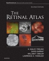
    The Retinal Atlas

    AvK. Bailey Freund,David Sarraf

    Inbunden, Engelska, 2016

    3 309 kr

    Beställningsvara. Skickas inom 5-8 vardagar. Fri frakt över 249 kr.

    Beskrivning

    With more than 5,000 images, a unique page layout, and comprehensive illustrations of the entire spectrum of vitreous, retina, and macula disorders, The Retinal Atlas, 2nd Edition, is an indispensable reference for retina specialists and comprehensive ophthalmologists as well as residents and fellows in training. For this edition, an expanded author team made up of Drs. K. Bailey Freund, David Sarraf, William F. Mieler, and Lawrence A. Yannuzzi, each an expert in retinal research and imaging, provide definitive up-to-date perspectives in this rapidly advancing field. This award-winning title has been thoroughly updated with new images with multimodal illustrations, new coverage and insight into key topics, and new disorders and classifications, while retaining the innovative page layout that has made it the most useful and most complete atlas of its kind.

    Produktinformation

    • Utgivningsdatum:2016-11-30
    • Mått:224 x 282 x 56 mm
    • Vikt:3 496 g
    • Format:Inbunden
    • Språk:Engelska
    • Antal sidor:1 200
    • Upplaga:2
    • Förlag:Elsevier Health Sciences
    • ISBN:9780323287920

    Utforska kategorier

    • Oftalmologi inom Medicin

    Recensioner i media

    "a user-friendly and comprehensive retinal atlas""As a reference textbook predominantly consisting of images of retinal disease (and optic nerve disorders for good measure), this is a diagnostician’s dream.""an essential text for eye care professionals ensuring timely diagnosis of retinal disease""a pleasure to browse, visually attractive, easy to navigate and an encyclopaedia of retinal disease" --2017 BMA Awards: Highly Commended, Internal MedicinePraise for the previous edition..."Lawrence Yannuzzi has undertaken this colossal project and delivered a masterpiece… The Retinal Atlas is an outstanding book. It is an instant classic that I highly recommend to every retina specialist, retina fellow, and ophthalmology resident." Reviewed by JAMA. 2011;305(18):1915Yannuzzi's new book, The Retinal Atlas, is a 900-page atlas of heroic proportion, containing thousands of illustrative clinical images... The book is in an engaging and inviting format - images and clinical pearls on every page. This book belongs in every ophthalmology department's library and academic retinal specialists' office for educating residents and fellows. It would also be an invaluable reference book for any clinical practice that encounters out-of-the-ordinary retinal diseases. The ability to view the book online is especially helpful when the atlas is not nearby." Reviewed by: American Journal of Ophthalmology (Volume 151, Issue 2, February 2011)**2010 PROSE Award Winner for Clinical Medicine.**"For eye care students and professionals in any context, this reference offers many hundreds of captioned images, presented along with diagnostic commentary, illustrating a wide range of diseases and complications. Color-coded borders identify five categories of imaging: general, fluorescein angiogram, fundus autofluorescence, indocyanine green angiogram, and red free photograph. Material is arranged in 15 chapters beginning with the normal retina and continuing with hereditary chorieoretinal dystrophies, the pediatric retina, inflammation, infection, retinal vascular disease, degeneration, oncology, macular fibrosis (and pucker, cysts, holes, folds, and edema), non-rhegmatogenous retinal detachment, peripheral degenerations, trauma, surgery complications, toxicities, and congenital anomalies of the optic nerve. Yannuzzi (Manhattan Eye Ear Throat Hospital, and Columbia U. Medical School) is credited as author, but he acknowledges contributions from nine other doctors who have helped in their areas of expertise. Purchase of the text includes access to the contents online." Reviewed by: Book News, Inc."This book is an excellent, visually appealing and comprehensive resource...well-written and enhance the reader’s experience and understanding" Reviewed by The South African Optometrist, November 2010

    Innehållsförteckning

    • 1. Normal2. Hereditary chorioretinal dystrophies3. Pediatric retina4. Inflammation5. Infection6. Retinal vascular disease7. Degeneration8. Oncology9. Macular fibrosis, pucker, cysts, holes, folds, and edema10. Non-rhegmatogenous retinal detachment11. Peripheral retinal degenerations and rhegmatogenous retinal detachment12. Traumatic chorioretinopathy13. Complications of ocular surgery14. Chorioretinal toxicities15. Congenital anomalies of the optic nerve