• Fri frakt över 249 kr
  • •
  • Snabba leveranser
  • •
  • Billiga böcker
Kundservice

Du är på sajten för privatpersoner.

Företag, bibliotek eller offentlig verksamhet?

Du handlar på classic.bokus.com, där alla dina funktioner finns intakta.
Till classic.bokus.com
Bokus logotyp. Gå till startsidan.
  • Erbjudanden
  • Nyheter
  • Student
  • Topplistor
  • Barn & ungdom
  • Bokus Play
  • E-böcker
  • Pocketböcker
  • Spel & pussel

10% rabatt på allt med kod: NYSTART10 →

Sidfot

Mina sidor

    Hjälp

    • Kundservice
    • Vanliga frågor och svar
    • Frakt och leverans
    • Retur vid ångerrätt
    • Reklamera vara
    • Betalning
    • Köpvillkor
    • Allmänna villkor
    • Information om webbplatsens tillgänglighet

    Om Bokus

    • Om oss
    • Pressrum
    • För studenter
    • För företag
    • För bibliotek och offentlig verksamhet
    • För leverantörer
    • Hållbarhet

    Populärt

    • Aktuella erbjudanden
    • Presentkort
    • Studentlitteratur
    • Nya böcker
    • Topplistor
    • Signerade böcker
    • Engelska böcker

    Inspiration

    • Boktips
    • BookTok
    • Populära bokserier
    • Barnbokskaraktärer
    • Populära författare
    Logotyp för Bokus
    Följ oss på Facebook (extern länk)Följ oss på Instagram (extern länk)Följ oss på YouTube (extern länk)Följ oss på TikTok (extern länk)
    bokus @ CookiesAnpassa cookiesIntegritetspolicyKöpvillkor
    Till Citymail hemsida (extern länk)Till Budbee hemsida (extern länk)Till Postnord hemsida (extern länk)Till Schenker hemsida (extern länk)Till Early Bird hemsida (extern länk)Till Walleys hemsida (extern länk)
    1. Medicin
    2. Andra medicinska specialiteter
    3. Patologi

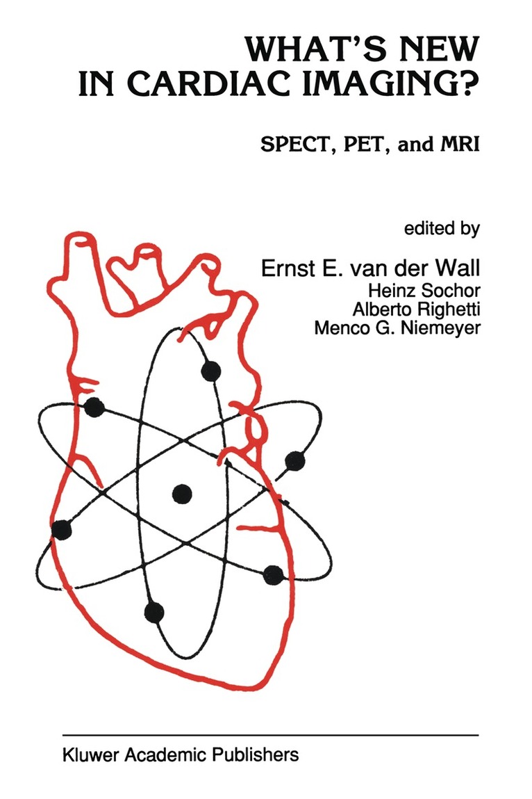
    What’s New in Cardiac Imaging?

    SPECT, PET, and MRI

    AvE. Van Der Wall,Ernst E. van der Wall

    Inbunden, Engelska, 1992

    Del i serien Developments in Cardiovascular Medicine

    3 340 kr

    Beställningsvara. Skickas inom 10-15 vardagar. Fri frakt över 249 kr.

    Beskrivning

    Since the introduction of myocardial perfusion imaging and radionuclide angiography in the mid-seventies, cardiovascular nuclear medicine has undergone an explosive growth. The use of nuclear cardiology techniques has become one of the cornerstones of the noninvasive assessment of coronary artery disease. In the past 15 years major steps have been made from visual analysis to quantitative analysis, from planar imaging to tomographic imaging, from detection of disease to prognosis, and from separate evaluations of perfusion, metabolism, and function to an integrated assessment of myocardial viability. In recent years many more advances have been made in cardiovascular nuclear imaging, such as the development of new imaging agents, reevaluation of existing procedures, and new clinical applications. This book describes the most recent developments in nuclear cardiology and also addresses new contrast agents in MRI. What's New in Cardiac Imaging will assist the clinical cardiologist, the cardiology fellow, the nuclear medicine physician, and the radiologist in understanding the most recent achievements in clinical cardiovascular nuclear imaging.

    Produktinformation

    • Utgivningsdatum:1992-07-31
    • Mått:155 x 235 x 41 mm
    • Vikt:1 160 g
    • Format:Inbunden
    • Språk:Engelska
    • Serie:Developments in Cardiovascular Medicine
    • Antal sidor:550
    • Upplaga:1992
    • Förlag:Kluwer Academic Publishers
    • ISBN:9780792316152

    Utforska kategorier

    • Patologi inom Medicin
    • Hjärt- och kärlsjukdomar inom Medicin
    • Medicinsk bildbehandling inom Medicin

    Innehållsförteckning

    • 1. What’s new in cardiac imaging?.- 2. Why new cardiac imaging agents?.- Section one: Perfusion.- 3. The value of measuring myocardial perfusion in coronary artery disease.- Single photon imaging.- 4. Myocardial perfusion imaging with xenon-133.- 5. Myocardial perfusion and krypton-81m.- 6. Myocardial perfusion imaging with technetium-99m isonitriles; attractive thallium substitutes?.- 7. Is there a specific role for technetium-99m SestaMIBI in the assessment of cardiac arrhythmias?.- 8. Technetium-99m complexes of functionalized diphosphines for myocardial perfusion imaging in man.- 9. Myocardial perfusion imaging with technetium-99m teboroxime.- Positron emission tomography.- 10. Clinical applications of rubidium-82 myocardial perfusion imaging.- 11. Nitrogen-13 ammonia perfusion imaging.- 12. Quantification of myocardial perfusion with oxygen-15 water.- 13. Myocardial perfusion imaging with copper-62 labeled Cu-PTSM.- Magnetic resonance imaging.- 14. Application of contrast agents in magnetic resonance imaging: additional value for detection of myocardial ischemia?.- 15. Magnetic resonance imaging using paramagnetic contrast agents in the clinical evaluation of myocardial infarction.- Section two: Metabolism.- 16. Assessment of myocardial metabolism in vivo. a biochemist’ view.- Single photon imaging.- 17. Myocardial metabolic imaging with iodine-123 fatty acids.- Positron emission tomography.- 18. Assessment of myocardial fatty acid metabolism with carbon-11 palmitate.- 19. Myocardial metabolic imaging with fluorine-18 deoxyglucose.- 20. Myocardial metabolic imaging with carbon-11-acetate.- Section three: Infarct-avid imaging.- Single photon imaging.- 21. Infarct-avid imaging: usefulness, problems and limitations.- 22. Myocyte necrosis-avid with radiolabeledantimyosin antibody: experimental and clinical acute myocardial infarction, myocarditis and heart transplant rejection.- 23. Myocardial infarct imaging with cardiac troponin-I antibodies.- Section four: Function.- Single photon imaging, positron emission tomography.- 24. Radionuclide imaging in the evaluation of cardiac function: new developments?.- Single photon imaging.- 25. Krypton-81m equilibrium radionuclide ventriculography for the assessment of right ventricular function.- 26. Simultaneous assessment of myocardial function and perfusion.- Section five: Sympathetic nerve system.- Single photon imaging.- 27. Radiolabeled metaiodobenzylguanidine (1-123 MIBG): value in clinical cardiology?.- 28. Scintigraphic assessment of cardiac innervation using iodine-123 metaiodobenzylguanidine.- 29. Clinical experience with iodine-123 metaiodobenzylguanidine.- Positron emission tomography.- 30. Studies of cardiac receptors by positron emission tomography.- 31. Heart neuronal imaging with carbon-11- and fluorine-18-labeled tracers.- Section six: Leukocytes, platelets, lipoproteins.- Single photon imaging.- 32. Labeling of leukocytes, platelets, and lipoproteins: useful in clinical cardiology?.- 33. Indium-111 leukocyte scintigraphy for detection of valvular abscesses and vegetations.- 34. Detection of cardiac thrombi with indium-111 platelet scintigraphy.- 35. Scintigraphic detection of atherosclerosis with radiolabeled low density lipoprotein.- Section seven: Viability.- Single photon imaging, positron emission tomography, magnetic resonance imaging.- 36. Assessment of myocardial viability with scintigraphic techniques and magnetic resonance imaging: new attainments?.- Positron emission tomography.- 37. Is it worth assessing regional myocardial viability with positron emissiontomography?.- 38. Assessment of tissue viability after myocardial infarction with fluorine-18 deoxyglucose using planar imaging; the alternative approach.- Section eight: Alternative stress imaging.- Single photon imaging, echocardiography, magnetic resonance imaging.- 39. New developments in pharmacological stress imaging.