• Fri frakt över 249 kr
  • •
  • Snabba leveranser
  • •
  • Billiga böcker
Kundservice

Du är på sajten för privatpersoner.

Företag, bibliotek eller offentlig verksamhet?

Du handlar på classic.bokus.com, där alla dina funktioner finns intakta.
Till classic.bokus.com
Bokus logotyp. Gå till startsidan.
  • Erbjudanden
  • Nyheter
  • Student
  • Topplistor
  • Barn & ungdom
  • Bokus Play
  • E-böcker
  • Pocketböcker
  • Spel & pussel

10% rabatt på allt med kod NYSTART10 →

Sidfot

Mina sidor

    Hjälp

    • Kundservice
    • Vanliga frågor och svar
    • Frakt och leverans
    • Retur vid ångerrätt
    • Reklamera vara
    • Betalning
    • Köpvillkor
    • Allmänna villkor
    • Information om webbplatsens tillgänglighet

    Om Bokus

    • Om oss
    • Pressrum
    • För studenter
    • För företag
    • För bibliotek och offentlig verksamhet
    • För leverantörer
    • Hållbarhet

    Populärt

    • Aktuella erbjudanden
    • Presentkort
    • Studentlitteratur
    • Nya böcker
    • Topplistor
    • Signerade böcker
    • Engelska böcker

    Inspiration

    • Boktips
    • BookTok
    • Populära bokserier
    • Barnbokskaraktärer
    • Populära författare
    Logotyp för Bokus
    Följ oss på Facebook (extern länk)Följ oss på Instagram (extern länk)Följ oss på YouTube (extern länk)Följ oss på TikTok (extern länk)
    bokus @ CookiesAnpassa cookiesIntegritetspolicyKöpvillkor
    Till Citymail hemsida (extern länk)Till Budbee hemsida (extern länk)Till Postnord hemsida (extern länk)Till Schenker hemsida (extern länk)Till Early Bird hemsida (extern länk)Till Walleys hemsida (extern länk)
    1. Naturvetenskap och teknik
    2. Matematik och naturvetenskap
    3. Biologi

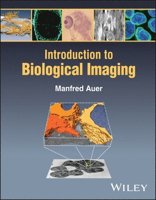
    Introduction to Biological Imaging

    AvManfred Auer

    Häftad, Engelska, 2024

    1 616 kr

    Beställningsvara. Skickas inom 5-8 vardagar. Fri frakt över 249 kr.

    Fler format och utgåvor

    E-bok

    1 819 kr

    E-bok

    1 814 kr

    Beskrivning

    Discover what biological imaging is able to accomplish in this up-to-date textbook One of the fundamental goals of biology is to understand how living organisms establish and maintain their spatiotemporal organization of the biochemical, cell biological and developmental biology processes that sustain life. Biological systems are inherently complex with a large number of components needed to sustain cellular function. In order to understand any complex system, one must determine its composition by identifying the components it is made of, how each of these components function and carry out their specific task, and how they interact with one another to function together. To grasp the link of such changes to physiological cell and tissue function and/or pathogenesis/disease progression, we need to understand how modifications alter macromolecular function, macromolecular interactions, and/or spatiotemporal distribution and overall supramolecular structural organization. Biological imaging holds the key to understanding spatiotemporal organization, and will thus be increasingly important for the next generations of biological and biochemical researchers. Introduction to Biological Imaging provides the first comprehensive textbook surveying this subject. It elucidates the fundamental principles underlying the capture and production of bioimages, the requirements of image analysis and interpretation, and some key problems and solutions in bioimaging. It includes everything experimental biologists need to incorporate appropriate bioimaging solutions into their work. Introduction to Biological Imaging readers will also find: Coverage of all major types of biological imaging, including medical imaging, cellular imaging, macromolecular imaging, and moreAdvice on preparing samples for various imaging methodsSpecific examples in each chapter connecting bioimaging process to the production of real experimental dataIntroduction to Biological Imaging is a valuable introduction for undergraduate or graduate students in courses relating to bioimaging, as well as scientists and researchers in the biological and medical fields who want a one-stop reference for the full range of imaging techniques.

    Produktinformation

    • Utgivningsdatum:2024-05-02
    • Mått:216 x 274 x 18 mm
    • Vikt:953 g
    • Format:Häftad
    • Språk:Engelska
    • Antal sidor:352
    • Förlag:John Wiley & Sons Inc
    • ISBN:9781119705949

    Utforska kategorier

    • Biologi inom Naturvetenskap och teknik
    • Medicinsk bildbehandling inom Medicin

    Mer om författaren

    Manfred Auer, PhD, is Professor in the School of Biological Sciences and Medical Engineering at Southeast University in Nanjing, China. He has decades of research experience in Europe and the US, and is the recipient of honors including an Otto Hahn Award of the Max Planck Society for exceptional PhD thesis (1999), an U.S. Department of Energy Outstanding Mentoring Award (2009), a Berkeley Lab Prize (2012), and a Chinese Ministry of Education Chang Jiang Scholar Chair Professor Award (2022).

    Innehållsförteckning

    • Preface xiii1 Introduction – A Brief History of Imaging 11.1 Spatiotemporally Organization of Biological Processes 21.1.1 Metabolism 21.1.2 Developmental Biology 21.1.3 Disease Mechanisms 21.2 Why Imaging? 31.2.1 A Very Brief History of Cellular Biological Imaging 31.2.2 A Very Brief History of Macromolecular Biological Imaging 52 Basics of Atomic and Molecular Physics 92.1 Matter and Energy 92.1.1 Standard Model of Particle Physics 92.1.2 Atomic Structure 112.1.3 Periodic Table of Elements 162.1.4 Transitions of Electrons 172.1.5 Molecular Orbitals 172.1.6 Forces and Bonds Between Neighboring Atoms 182.2 Electromagnetic Spectrum 212.2.1 Gamma Rays 222.2.2 X-Rays 222.2.3 Ultraviolet Radiation 222.2.4 Visible Light 222.2.5 Infrared Radiation 232.2.6 Microwave Radiation 242.2.7 Radio Wave Radiation 242.3 Interaction of Radiation with Matter 242.3.1 High-Energy Photons 242.3.2 High-Energy Particles 252.3.3 Middle-Energy Photons 252.3.4 Low-Energy Photons 263 Basics of Optics 333.1 Properties of Light 333.2 Classical Optics 333.3 Interaction of Light with Matter 343.3.1 Reflection 343.3.2 Refraction 343.3.3 Dispersion 353.4 Lenses 353.5 Numerical Aperture 353.6 Scattering 363.7 Interference 373.8 Diffraction 373.9 Airy Disk 393.10 Point-Spread Function 393.11 Resolution 403.12 Aberrations 413.13 Polarization 413.14 Photoelectric Effect 423.15 Valence Electron Transitions 423.16 Vibrational and Rotational Transitions 433.17 Stimulated Emission 434 Basics of Fourier “Math” 455 Basics of Imaging and Image Formation 515.1 Goals of Imaging 515.2 Source of Radiation 525.2.1 Gamma-Rays 525.2.2 X-Rays 535.2.3 UV-Rays/Visible Light Rays/Near-IR Light Rays 535.2.4 IR Radiation 555.2.5 Microwave Radiation 555.2.6 Radio Wave Radiation 555.2.7 Particle Radiation 555.2.8 Particle Radiation: Electrons 565.2.9 Particle Radiation: Positrons 575.2.10 Particle Radiation: Alpha-Radiation/Helium Nuclei 575.2.11 Particle Radiation: Ion Beams 575.2.12 Particle Radiation: Neutrons 575.2.13 Particle Radiation: Protons 575.3 Beam Collimation 575.4 Lenses 585.5 Lens-Free Imaging/Near Field Imaging 595.6 Signal Detection and Detectors 605.6.1 What Are the Different Types of Radiation That Need to Be Detected? 605.6.2 Detection of Particle Radiation 615.6.3 Detection of Ionizing Electromagnetic Radiation 625.6.4 Detection of Non-Ionizing Electromagnetic Radiation 635.6.5 Principles of Signal Detection 645.7 Modulation-, Contrast-, and Optical-Transfer Function 695.8 Field of View, Coverage 705.9 Spot-Scan Versus Flood-Beam Imaging 715.10 Radiation Damage 725.11 Spectroscopic Imaging 725.12 Multidimensional and Multi-channel Imaging 735.13 Sample Preparation Requirements 755.13.1 Macromolecular 3D Structure 755.13.2 Subcellular, Cellular, and Tissue 3D Architecture 765.13.3 Dynamic Localization of Macromolecules in Cells and Tissues 775.13.4 Chemical Inventory of Cells and Tissues 775.13.5 Medical Imaging of Tissues and Organs 785.14 Labels and Labeling 795.15 Label-Free Imaging 815.16 2D Versus 3D Imaging 825.17 Structural Versus Functional Imaging 855.18 Correlative Imaging 856 Basics of Image Processing and Image Analysis 876.1 Overview 876.2 Characteristics of Digital Images 896.3 Compression of Image Data 966.4 Metadata 986.5 Spatial, Temporal and Spectral Resolution 996.6 Image Intensity Values 996.6.1 Contrast 996.6.2 Histogram 1006.6.3 False-Color Representation and Lookup Tables 1016.7 Raster Graphics Versus Vector Graphics 1016.8 Simple Image Processing Operations 1026.9 Image Restoration 1046.10 Image Enhancement 1046.11 Image Reconstruction 1096.12 Visualization 1096.12.1 Introduction to Scientific Visualization 1096.12.2 Visualization Software Examples 1136.12.3 3D Volume Image Data 1146.12.4 Visualizing Macromolecular Imaging Data 1156.12.5 Visualizing Cellular Imaging Data 1166.12.6 A Few More Points Worth Noting 1196.13 Image Segmentation 1196.13.1 Map Density Value- Based Segmentation 1206.13.2 Manual Segmentation – Creating Regions of Interest 1216.13.3 Automated Segmentation 1226.14 Making Changes to Object Selection: Dilation, Erosion, Closing, Filling Holes 1316.15 Measurements/Quantitative Analysis 1317 Techniques – Macromolecular Structure Determination 1357.1 X-Ray Crystallography 1357.1.1 Overview 1357.1.2 Historical Perspective/Milestones 1377.1.3 Theoretical Background 1387.1.4 Instrumentation/Experimental Setup 1437.1.5 Data Collection 1467.1.6 Data Analysis and Phasing 1477.1.7 Map Calculation and Model Building 1487.1.8 Refinement and Validation 1487.1.9 Visualization 1497.1.10 Outlook 1497.2 Macromolecular Cryo-Electron Microscopy 1507.2.1 Overview 1507.2.2 Historical Perspective/Milestones 1527.2.3 Theoretical Background 1627.2.4 Instrumentation/Experimental Setup 1657.2.5 Sample Preparation and Data Collection 1687.2.6 Data Analysis 1697.2.7 Map Calculation and Resolution Estimates 1757.2.8 Visualization and Model Building 1767.3 NMR Spectroscopy 1777.3.1 Overview 1777.3.2 Historical Perspective/Milestones 1787.3.3 Theoretical Background 1797.3.4 Instrumentation and Experimental Setup 1817.3.5 Data Collection and Analysis 1817.4 Small Angle Scattering – SAXS and SANS 1847.4.1 Small-Angle X-Ray Scattering (SAXS) 1847.4.2 Small-Angle Neutron Scattering (SANS) 1847.5 Atomic Force Microscopy (AFM) 1858 Techniques – Cell and Tissue Architectural Imaging 1898.1 Overview 1898.2 Historical Perspective/Milestones 1918.2.1 Transmission Electron Microscopy (TEM) Imaging 1958.2.2 Scanning Electron Microscopy (SEM) Imaging 1968.2.3 Helium Ion Microscopy Imaging 1968.2.4 X-Ray Microscopy Tomography Imaging 1978.2.5 Optical/Fluorescence Light Microscopy Imaging 1998.3 Theoretical Background 1998.3.1 Contrast Generation in Transmission Electron Microscopy 1998.3.2 Contrast Generation in Scanning Electron Microscopy (SEM) 2008.3.3 Contrast Generation in Helium Ion Microscopy (HeIM) 2018.3.4 Contrast Generation in X-Ray Microscopy Tomography (XRMT) 2018.3.5 3D Reconstructions of Pleiomorphic Objects 2038.4 Sample Preparation 2058.4.1 Electron Microcopy 2058.4.2 Need for Thin Samples 2058.4.3 Conventional TEM Sample Preparation 2058.4.4 Cryogenic EM Sample Preparation 2068.4.5 Sample Preparation for cryo-XRMT 2088.5 Instrumentation 2088.5.1 Radiation Source 2098.5.2 Electron/Ion/X-Ray Optical System 2108.5.3 Detectors 2118.5.4 Specimen Stage 2118.5.5 Vacuum System 2138.6 Data Collection and 3D Reconstruction 2148.6.1 Serial Section TEM 2148.6.2 Serial Section SEM 2158.6.3 Serial Block Face SEM 2158.6.4 Focused Ion Beam (Dual Beam) SEM 2158.6.5 X-Ray Microscopy Tomography 2168.7 Data Visualization and Analysis 2178.7.1 Visualization of Cellular Sceneries 2188.7.2 Segmentation/Feature Extraction 2198.7.3 Model Building and (Quantitative) Analysis 2218.7.4 Deriving Biological Meaning Through Interpretation 2229 Techniques – Cell and Tissue Characterization and Localization Imaging 2259.1 Overview 2259.2 Historical Perspective/Milestones 2269.2.1 What Is Light? 2269.2.2 Optical Instruments and Microscopy 2289.2.3 Application of Microscopy to Biological Samples 2309.2.4 Fluorescence Microscopy 2319.3 Theoretical Background 2329.4 Instrumentation/Experimental Setup 2359.4.1 Frame, Stage, and Sample Holders 2359.4.2 Light Source and Illumination 2379.4.3 Optical Path 2389.4.4 Detector 2399.5 Data Collection 2419.5.1 Choosing the Right Microscopy Technique 2419.5.2 White Light Imaging 2419.5.3 Fluorescence Microscopy 2439.6 Fluorophore Considerations 2449.6.1 Synthetic Organic Dyes 2449.6.2 Quantum Dots 2469.6.3 Immuno-Affinity-Based Labeling 2469.7 Genetically Encoded Fluorescent Proteins 2479.7.1 Intrinsically Fluorescent Proteins 2479.7.2 Photoactivatable Fluorescent Proteins 2489.7.3 Photoconvertible Fluorescent Proteins 2489.7.4 Photoswitchable Fluorescent Proteins 2489.7.5 Extrinsically Fluorescent Proteins 2489.8 RNA/DNA – Fluorescent Probes 2489.9 Fluorescent Imaging Types 2509.10 Detangling the Many Fluorescence Imaging Approaches 2509.11 Improve Axial Resolution and Reduce Phototoxicity 2519.11.1 Epifluorescence Microscopy 2519.11.2 Deconvolution Microscopy 2519.11.3 Confocal Laser Scanning Microscopy 2529.11.4 Spinning Disk Confocal Microscope 2529.11.5 4Pi and Confocal Theta Microscopy 2529.11.6 Two-Photon Microscopy 2539.11.7 Single Plane Illumination Microscopy (SPIM) 2559.11.8 Total-Internal Reflection Fluorescence (TIRF) Microscopy 2569.12 Superresolution Imaging – Breaking the Resolution Barrier 2579.12.1 Photoactivation 2579.12.2 Photoconversion 2579.12.3 Photoswitching 2589.13 Superresolution Imaging 2589.13.1 Single-Molecule Localization Microscopy 2589.13.2 Patterned Illumination Microscopy 2609.14 Near-Field Scanning Microscopy 2619.15 Molecular Dynamics Study of Macromolecules 2619.15.1 Fluorescence Recovery After Photobleaching (FRAP) Microscopy 2619.15.2 Fluorescence Loss in Photobleaching (FLIP) Microscopy 2629.15.3 Fluorescence Speckle Microscopy (FSM) 2629.15.4 Förster Resonance Energy Transfer Microscopy 2629.15.5 Fluorescence Lifetime Imaging Microscopy 2649.16 Visualization 2649.17 Data Analysis 26510 Techniques – Chemical Imaging at the Cell and Tissue Level 26710.1 X-Ray Microanalysis: X-Ray Fluorescence Microscopy (XFM), Energy-Dispersive X-Ray Spectroscopy (EDX), Wavelength-Dispersive X-ray Spectroscopy (WDS) 26910.2 Raman Imaging 27110.2.1 Overview 27110.2.2 Theoretical Background 27210.2.3 Experimental Design 27610.2.4 Data Analysis 27610.3 Fourier-Transform Infrared (FTIR) Imaging 27710.4 Mass Spectrometry 27910.4.1 Overview 27910.4.2 Historical Perspective 28210.4.3 Instrumentation 28310.4.4 Data Analysis 28911 Techniques – Tissue and Organ Medical Imaging 29111.1 Positron Emission Tomography 29211.2 Computed (Axial) Tomography 29411.3 Magnetic Resonance Imaging (MRI) 29411.4 Medical Ultrasound 29611.5 Bioluminescence 29711.6 Optical Coherence Tomography 29711.7 Histology/Histopathology 29812 The Future of Bioimaging 30112.1 Future of Macromolecular Imaging 30112.2 Future of (Sub)Cellular and Tissue Imaging 30312.2.1 Light Microscopy Versus Electron Microscopy 30312.2.2 Cryo-Preservation in EM Sample Preparation 30412.2.3 FIB Milling Sample Preparation 30412.2.4 Electron Tomography 30412.2.5 Correlative Light and Electron Microscopy 30512.2.6 Volume-SEM 30512.2.7 Helium Ion Microscopy 30512.2.8 Cryo-Soft-X-Ray Microscopy Tomography 30612.2.9 Hard-X-Ray Microscopy Tomography 30612.3 Future of Chemical Imaging 30712.4 Future of Medical Imaging 30712.5 Future of Image Processing and Analysis 30812.6 Future of Multiscale Imaging 31012.7 Future of Multimodal Imaging – Correlative Imaging 31312.8 Future of Information Integration – Multiscale, Multimodal, and Integrated 315Index 319