E-bok, Engelska, 2011
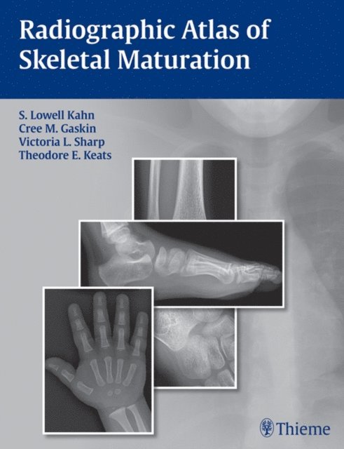
Radiographic Atlas of Skeletal Maturation
Av Bing Li, Theodore E. Keats, Victoria L. Sharp, Christopher M. Gaskin, S. Lowell Kahn
4279 kr
Läs direkt i Bokus Reader – eller ladda ned till din enhet
Beskrivning
Nearly 2,300 images provide the reference standard for normal skeletal maturation at every developmental stage
When dealing with the maturing skeleton and its many complex growth alterations, physicians are constantly faced with the question: "Is this image normal?" The Radiographic Atlas of Skeletal Maturation succinctly answers that question by providing a comprehensive set of male and female reference images for every age and body part. This allows physicians to quickly hone in on "normal" ranges for the specific case they are reviewing--particularly useful when called upon to read a pediatric skeletal radiograph in the emergency room or while on call.
Special Features:
- Access to nearly 2,300 high-quality images that provide instant reference to "normal" views of the skeleton at every developmental milestone-available in both the text and accompanying DVD
- Multiple projections at every age, sex, and body part combination so that the user can match the reference points in the book to the case at hand and arrive at a solid clinical interpretation
- Practical text layout organized by gender and body part that provides quick access to images of normal development at any given age
- A software virtual "skeletal survey" demonstrates images of younger and older individuals and crystallizes the subtle variations in growth patterns
- Powerful software package with advanced image enhancement tools allows optimization of atlas image details for greater clarity. Compatible with numerous image formats (including DICOM) allowing viewing and editing of outside images
- Convenient growth charts included in the book and DVD
This unique resource, with its vast collection of print and DVD images of normal progressive skeletal development, gives physicians the full range of comparative information they need to interpret pediatric skeletal radiographs in any clinical setting. It is the reference standard for radiologists, pediatricians, orthopedists, emergency room physicians, internists, rehabilitation physicians, and training physicians who are called upon to review a pediatric radiograph and confidently make a diagnosis.
Produktinformation
- Utgivningsdatum: 2011-11-22
- Språk: Engelska
- Filformat: EPUB
- Kopieringsskydd: LCP
- ISBN: 9781638532217
- Förlag: Thieme Publishers New York
Utforska kategorier
Betyg & recensioner
0 recensioner
Inga recensioner tillgängliga.