• Fri frakt över 249 kr
  • •
  • Snabba leveranser
  • •
  • Billiga böcker
Kundservice

Du är på sajten för privatpersoner.

Företag, bibliotek eller offentlig verksamhet?

Du handlar på classic.bokus.com, där alla dina funktioner finns intakta.
Till classic.bokus.com
Bokus logotyp. Gå till startsidan.
  • Erbjudanden
  • Nyheter
  • Student
  • Topplistor
  • Barn & ungdom
  • Bokus Play
  • E-böcker
  • Pocketböcker
  • Spel & pussel

10% rabatt på allt med kod: NYSTART10 →

Sidfot

Mina sidor

    Hjälp

    • Kundservice
    • Vanliga frågor och svar
    • Frakt och leverans
    • Retur vid ångerrätt
    • Reklamera vara
    • Betalning
    • Köpvillkor
    • Allmänna villkor
    • Information om webbplatsens tillgänglighet

    Om Bokus

    • Om oss
    • Pressrum
    • För studenter
    • För företag
    • För bibliotek och offentlig verksamhet
    • För leverantörer
    • Hållbarhet

    Populärt

    • Aktuella erbjudanden
    • Presentkort
    • Studentlitteratur
    • Nya böcker
    • Topplistor
    • Signerade böcker
    • Engelska böcker

    Inspiration

    • Boktips
    • BookTok
    • Populära bokserier
    • Barnbokskaraktärer
    • Populära författare
    Logotyp för Bokus
    Följ oss på Facebook (extern länk)Följ oss på Instagram (extern länk)Följ oss på YouTube (extern länk)Följ oss på TikTok (extern länk)
    bokus @ CookiesAnpassa cookiesIntegritetspolicyKöpvillkor
    Till Citymail hemsida (extern länk)Till Budbee hemsida (extern länk)Till Postnord hemsida (extern länk)Till Schenker hemsida (extern länk)Till Early Bird hemsida (extern länk)Till Walleys hemsida (extern länk)
    1. Naturvetenskap och teknik
    2. Matematik och naturvetenskap
    3. Matematik

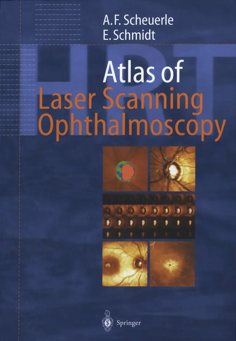
    Atlas of Laser Scanning Ophthalmoscopy

    AvAlexander Friedrich Scheuerle,Eckart Schmidt

    Häftad, Engelska, 2012

    563 kr

    Beställningsvara. Skickas inom 10-15 vardagar. Fri frakt över 249 kr.

    Fler format och utgåvor

    Inbunden

    1 119 kr

    Beskrivning

    Glaucoma remains one of the leading causes of blindness. Laser scanning tomography has gained an indispensable role in the ophthalmologic diagnosis, especially in the long-term follow-up of glaucoma. Confocal laser scanning ophthalmoscopy provides key insights into the three-dimensional anatomy of the optic disc in vivo. This unique atlas contains superb images of all clinically relevant diseases diagnosed by current models of the Heidelberg Retina Tomograph. It correlates classical diagnostic tools like perimetry and fundus photography with state-of-the-art studies including digital retinal angiography, optical coherence tomography and laser scanning tomography. Special features include the illustrated coverage of:Diseases of the optic nerve head; different types and stages of glaucoma, non-glaucomatous neuropathy and papilledema; automated classification procedures for the detection of glaucoma; strategies for the interpretation of follow-up results in optic disc monitoring.Macular diseases: the shortly released Retina Module expands the diagnostic spectrum of laser scanning ophthalmoscopy significantly, adding measuring and monitoring of diabetic and cystoid macular edema.This atlas is the most comprehensive up-to-date reference of laser scanning ophthalmoscopy available, ideal for residents and general ophthalmologists who want to enhance their diagnostic skills.

    Produktinformation

    • Utgivningsdatum:2012-01-19
    • Mått:193 x 260 x 11 mm
    • Vikt:413 g
    • Format:Häftad
    • Språk:Engelska
    • Antal sidor:170
    • Upplaga:2004
    • Förlag:Springer-Verlag Berlin and Heidelberg GmbH & Co. KG
    • ISBN:9783642639210

    Utforska kategorier

    • Matematik inom Naturvetenskap och teknik
    • Oftalmologi inom Medicin

    Innehållsförteckning

    • 1 Introduction.- 1.1 Why Is Laser Scanning Ophthalmoscopy Needed?.- 1.2 Technical Background.- 1.3 Stereometric Parameters of the HRT.- 2 Healthy Optic Discs.- 2.1 Natural Variety of Optic Discs.- 2.2 Regular Shapes.- 2.3 Micropapillas.- 2.4 Megalopapillas.- 2.5 Tilted Discs.- 3 Glaucomas.- 3.1 Automated Classification Procedures.- 3.2 Moderate Glaucomas.- 3.3 Nerve Fiber Bundle Defects.- 3.4 Advanced Glaucomas.- 3.5 Micropapillary Glaucomas.- 3.6 Megalopapillary Glaucomas.- 3.7 Normal Tension Glaucomas.- 3.8 Classification Errors.- 4 Follow-up Examinations.- 4.1 Strategies for Longitudinal Analysis.- 4.2 Progression of Glaucoma.- 5 Prominent Optic Discs.- 5.1 Scanning of Prominent Discs.- 5.2 Drusen of the Optic Nerve.- 5.3 Optic Nerve Swelling.- 5.4 Papilledemas.- 6 Macular Scans.- 6.1 The Macular Edema Software Module.- 6.2 Cystoid Macular Edema.- 6.3 Branch Vein Occlusion.- 6.4 Central Serous Retinopathy.- 6.5 Macular Puckers.- 6.6 Macular Holes.- 7 Miscellaneous.- 7.1 Optimizing Imaging.- 7.2 Astigmatism.- 7.3 Retinal Irregularities.- 7.4 Floaters.- 8 Confocal Corneal Imaging.- 8.1 The Rostock Cornea Modu.- References.