• Fri frakt över 249 kr
  • •
  • Snabba leveranser
  • •
  • Billiga böcker
Kundservice

Du är på sajten för privatpersoner.

Företag, bibliotek eller offentlig verksamhet?

Du handlar på classic.bokus.com, där alla dina funktioner finns intakta.
Till classic.bokus.com
Bokus logotyp. Gå till startsidan.
  • Erbjudanden
  • Nyheter
  • Student
  • Topplistor
  • Barn & ungdom
  • Bokus Play
  • E-böcker
  • Pocketböcker
  • Spel & pussel

10% rabatt på allt med kod: NYSTART10 →

Sidfot

Mina sidor

    Hjälp

    • Kundservice
    • Vanliga frågor och svar
    • Frakt och leverans
    • Retur vid ångerrätt
    • Reklamera vara
    • Betalning
    • Köpvillkor
    • Allmänna villkor
    • Information om webbplatsens tillgänglighet

    Om Bokus

    • Om oss
    • Pressrum
    • För studenter
    • För företag
    • För bibliotek och offentlig verksamhet
    • För leverantörer
    • Hållbarhet

    Populärt

    • Aktuella erbjudanden
    • Presentkort
    • Studentlitteratur
    • Nya böcker
    • Topplistor
    • Signerade böcker
    • Engelska böcker

    Inspiration

    • Boktips
    • BookTok
    • Populära bokserier
    • Barnbokskaraktärer
    • Populära författare
    Logotyp för Bokus
    Följ oss på Facebook (extern länk)Följ oss på Instagram (extern länk)Följ oss på YouTube (extern länk)Följ oss på TikTok (extern länk)
    bokus @ CookiesAnpassa cookiesIntegritetspolicyKöpvillkor
    Till Citymail hemsida (extern länk)Till Budbee hemsida (extern länk)Till Postnord hemsida (extern länk)Till Schenker hemsida (extern länk)Till Early Bird hemsida (extern länk)Till Walleys hemsida (extern länk)
    1. Medicin
    2. Andra medicinska specialiteter
    3. Patologi

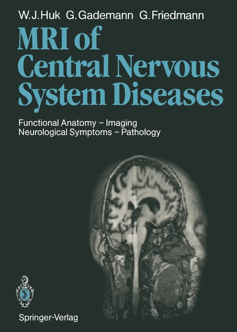
    Magnetic Resonance Imaging of Central Nervous System Diseases

    Functional Anatomy — Imaging Neurological Symptoms — Pathology

    AvWerner J. Huk,Günther F. Gademann

    Häftad, Engelska, 2011

    1 119 kr

    Beställningsvara. Skickas inom 10-15 vardagar. Fri frakt över 249 kr.

    Beskrivning

    Magnetic resonance imaging (MRI) is a new and still rapidly developing imaging technique which requires a new approach to image interpreta­ tion. Radiologists are compelled to translate their experience accumulat­ ed from X-ray techniques into the language of MRI, and likewise stu­ dents of radiology and interested clinicians need special training in both languages. Out of this necessity emerged the concept of this book as a manual on the application and evaluation of proton MRI for the radiolo­ gist and as a guide for the referring physician who wants to learn about the diagnostic value of MRI in specific conditions. After a short section on the basic principles of MRI, the contrast mechanisms of present-day imaging techniques, knowledge of which is essential for the analysis of relaxation times, are described in greater de­ tail. This is followed by a demonstration of functional neuroanatomy us­ ing three-dimensional view of MR images and a synopsis of frequent neurological symptoms and their topographic correlations, which will fa­ cilitate examination strategy with respect to both accurate diagnosis and economy.

    Produktinformation

    • Utgivningsdatum:2011-12-16
    • Mått:193 x 270 x 32 mm
    • Vikt:1 036 g
    • Format:Häftad
    • Språk:Engelska
    • Antal sidor:452
    • Förlag:Springer-Verlag Berlin and Heidelberg GmbH & Co. KG
    • ISBN:9783642725708
    • Översättare:Terry Telger

    Utforska kategorier

    • Patologi inom Medicin
    • Medicinsk bildbehandling inom Medicin
    • Neurokirurgi inom Medicin

    Innehållsförteckning

    • 1 Physical Principles and Techniques of MR Imaging.- 1.1 Physical Principles.- 1.2 Techniques of MR Imaging.- 1.3 Pulse Sequences and Image Contrasts.- 1.4 New Approaches in Clinical MR.- References.- 2 Normal Anatomy of the Central Nervous System.- 2.1 Orientation.- 2.2 Cerebral Cortex.- 2.3 Diencephalon and Basal Ganglia.- 2.4 Midbrain.- 2.5 Posterior Fossa.- 2.6 Optical System.- 2.7 Acoustic System.- 2.8 Cerebral Blood Supply.- 2.9 Spinal Cord.- References.- 3 Practical Aspects of the MR Examination.- 3.1 Preparations.- 3.2 Examination Procedure.- 3.3 Sedation, Anesthesia, and Anesthesiology Monitoring During MR Examinations.- 3.4 Side Effects and Contraindications.- References.- 4 Symptoms and Pathologic Anatomy: A Tabular Listing.- 5 General Aspects of the MR Signal Pattern of Certain Normal and Pathologic Structures.- 5.1 Brain Edema.- 5.2 Intracranial Hemorrhage and Iron Metabolism.- 5.3 Practical Aspects of Blood and CSF Flow.- 5.4 Effects of Radiotherapy on Brain and Spinal Cord Tumors.- 5.5 Effect of Contrast Agent (Gadolinium-DTPA) on the MR Appearance of Normal Tissue.- References.- 6 Magnetic Resonance Imaging of the Brain in Childhood: Development and Pathology.- 6.1 Introduction.- 6.2 Technical Aspects.- 6.3 Normal Appearance.- 6.4 Intracranial Hemorrhage.- 6.5 Infarction.- 6.6 Cysts and Leukomalacias.- 6.7 Hydrocephalus.- 6.8 Congenital Malformations.- 6.9 White Matter Disease.- 6.10 Infection.- 6.11 Delays or Deficits in Myelination.- 6.12 Tumors.- 6.13 Other Diseases.- 6.14 Follow-Up Examination.- 6.15 Conclusion.- References.- 7 Malformations of the CNS.- 7.1 Review of Ontogeny.- 7.2 MR Imaging of CNS Malformations.- 7.3 Midline Closure Defects (Neural Tube Defects, Dysraphic Disorders).- 7.4 Malformations of the Commissures and Midline Structures.- 7.5 Anomalies of Cell Migration.- 7.6 Destructive Anomalies.- 7.7 Neuroectodermal Dysplasias.- 7.8 Miscellaneous Abnormalities.- References.- 8 Degenerative Disorders of the Brain and White Matter Diseases.- 8.1 Primary Neuronal (Gray Matter) Involvement.- 8.2 Primary Myelin (White Matter) Involvement.- 8.3 Primary Involvement of the Basal Ganglia and Brainstem.- 8.4 Primary Involvement with No Sites of Predilection.- References.- 9 Temporal Lobe Epilepsy.- 9.1 MR Findings.- References.- 10 Intracranial Tumors.- 10.1 General Aspects of the Detection of Intracranial Tumors with MR.- 10.2 Tumors of Neuroepithelial Tissue.- 10.3 Tumors of the Nerve Sheath Cells.- 10.4 Tumors of Meningeal and Related Tissues.- 10.5 Primary Malignant Lymphomas.- 10.6 Tumors of Blood Vessel Origin.- 10.7 Germ Cell Tumors.- 10.8 Other Malformative Tumors and Tumor-Like Lesions.- 10.9 Tumors of the Pituitary.- 10.10 Local Extensions from Regional Tumors.- 10.11 Metastatic Tumors.- 10.12 Pseudotumor Cerebri.- References.- 11 Tumors of the Posterior Fossa.- 11.1 Intra-axial Tumors.- 11.2 Extra-axial Tumors.- References.- 12 Diseases of the Eyeball, Orbit, and Accessory Structures.- 12.1 Technical Requirements.- 12.2 Examination Technique.- 12.3 Eyeball.- 12.4 Optic Nerve.- 12.5 Lacrimal Glands.- 12.6 Ocular Muscles.- 12.7 Orbit.- 12.8 Malformations.- 12.9 Trauma.- References.- 13 Vascular Diseases of the Brain.- 13.1 Disturbances of Arterial Blood Flow.- 13.2 Disturbances of Venous Blood Flow.- 13.3 Hemorrhages.- References.- 14 Vascular Malformations.- 14.1 Vascular Malformations of the Brain.- 14.2 Vascular Malformations Involving the Vertebral Canal.- References.- 15 Inflammatory Diseases of the Central Nervous System.- 15.1 General Aspects of the Inflammatory Process.- 15.2 Classification.- 15.3 Infections of the Meninges.- 15.4 Infections of the Brain Tissue.- 15.5 Acquired Immune Deficiency Syndrome.- 15.6 Summary.- References.- 16 Head Injury.- 16.1 Intracranial Extracerebral Hemorrhages.- 16.2 Blunt Brain Injuries.- 16.3 Open Head Injuries.- 16.4 Other Sequelae of Injuries.- 16.5 Late Sequelae of Head Injuries.- References.- 17 Diseases of the Vertebral Column and Spinal Cord.- 17.1 Examination Technique.- 17.2 Intraspinal Processes.- 17.3 Spinal Tumors.- 17.4 Degenerative Diseases of the Spine.- 17.5 Trauma.- References.- Glossary of Magnetic Resonance Terms.