• Fri frakt över 249 kr
  • •
  • Snabba leveranser
  • •
  • Billiga böcker
Kundservice

Du är på sajten för privatpersoner.

Företag, bibliotek eller offentlig verksamhet?

Du handlar på classic.bokus.com, där alla dina funktioner finns intakta.
Till classic.bokus.com
Bokus logotyp. Gå till startsidan.
  • Erbjudanden
  • Nyheter
  • Student
  • Topplistor
  • Barn & ungdom
  • Bokus Play
  • E-böcker
  • Pocketböcker
  • Spel & pussel

10% rabatt på allt med kod: NYSTART10 →

Sidfot

Mina sidor

    Hjälp

    • Kundservice
    • Vanliga frågor och svar
    • Frakt och leverans
    • Retur vid ångerrätt
    • Reklamera vara
    • Betalning
    • Köpvillkor
    • Allmänna villkor
    • Information om webbplatsens tillgänglighet

    Om Bokus

    • Om oss
    • Pressrum
    • För studenter
    • För företag
    • För bibliotek och offentlig verksamhet
    • För leverantörer
    • Hållbarhet

    Populärt

    • Aktuella erbjudanden
    • Presentkort
    • Studentlitteratur
    • Nya böcker
    • Topplistor
    • Signerade böcker
    • Engelska böcker

    Inspiration

    • Boktips
    • BookTok
    • Populära bokserier
    • Barnbokskaraktärer
    • Populära författare
    Logotyp för Bokus
    Följ oss på Facebook (extern länk)Följ oss på Instagram (extern länk)Följ oss på YouTube (extern länk)Följ oss på TikTok (extern länk)
    bokus @ CookiesAnpassa cookiesIntegritetspolicyKöpvillkor
    Till Citymail hemsida (extern länk)Till Budbee hemsida (extern länk)Till Postnord hemsida (extern länk)Till Schenker hemsida (extern länk)Till Early Bird hemsida (extern länk)Till Walleys hemsida (extern länk)
    1. Medicin
    2. Andra medicinska specialiteter
    3. Medicinsk bildbehandling

    MRI of the Body

    AvDaniel Vanel,Michael T. McNamara

    Häftad, Engelska, 2012

    1 119 kr

    Beställningsvara. Skickas inom 10-15 vardagar. Fri frakt över 249 kr.

    Beskrivning

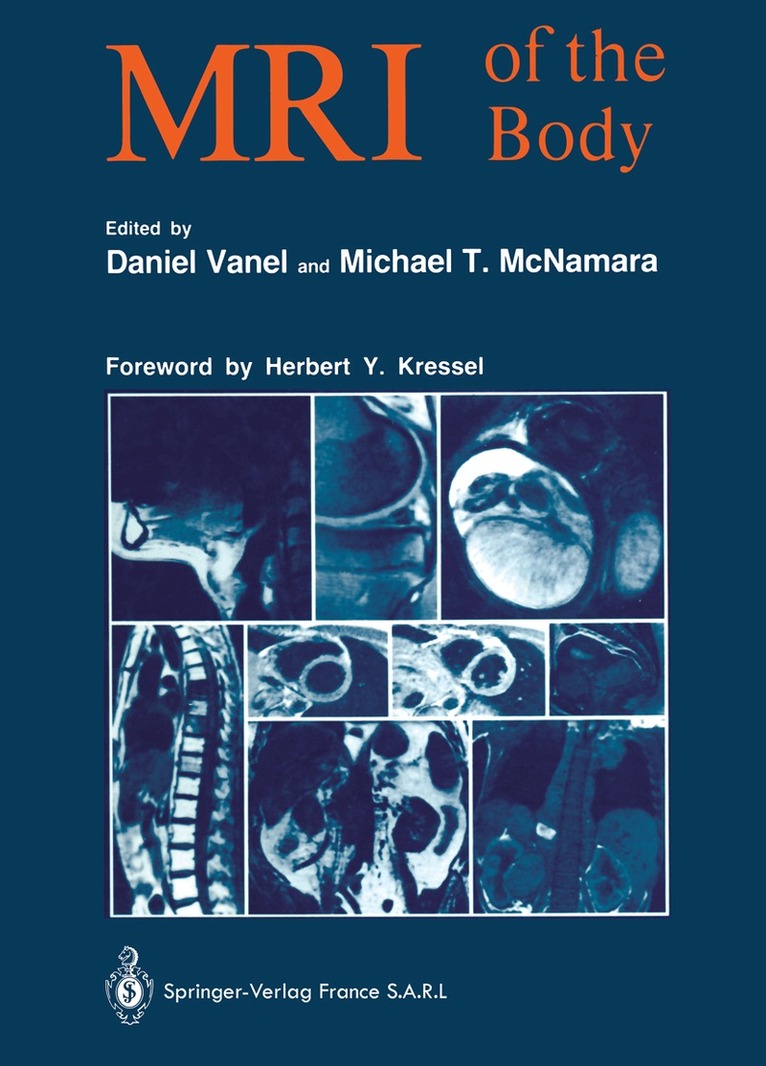
    As with the introduction of x -ray computed tomography, much of the initial development of magnetic resonance applications tended to focus on the central nervous system. The development of magnetic resonance imaging applications to other organ systems such as the chest, abdomen, pelvis and extremities has lagged somewhat behind, awaiting technical improvements, and a broader user base. The past two years have seen a marked increase in imaging applications throughout the body, most notably the musculoskeletal system. It is in this regard, that MRI of the Body is a welcome arrival as a text which describes both basic principles of magnetic resonance ima­ ging and surveys the current status of magnetic resonance imaging applications throughout the body. The volume is concise, focused, clinically oriented, and abun­ dantly illustrated. In each organ system, the appropriate technical approach is discussed, the normal anatomic features are reviewed, and the range of pathologic appearances which may be encountered are described. The authors of the chapters provide a balanced overview of MR applications and describe both present limitations and future potential of magnetic resonance imaging applications in the organ system described.

    Produktinformation

    • Utgivningsdatum:2012-08-17
    • Mått:210 x 279 x 23 mm
    • Vikt:999 g
    • Format:Häftad
    • Språk:Engelska
    • Antal sidor:387
    • Förlag:Springer-Verlag Berlin and Heidelberg GmbH & Co. KG
    • ISBN:9783642875588
    • Översättare:Susanne Assenat

    Utforska kategorier

    • Medicinsk bildbehandling inom Medicin

    Innehållsförteckning

    • Physical basis.- Physical basis of nuclear magnetic resonance.- Signal parameters.- Formation of an image.- Artifacts.- System-specific artifacts.- Patient-specific artifacts.- Quality control.- Definition of QC parameters.- Test substances and test objects.- NMR spectroscopy from experimental to clinical spectroscopy.- Principle of NMR spectroscopy.- Most significant results of spectrocopy in man.- Clinical application.- Conclusion.- Contrast media.- Theoretic basis.- Paramagnetic ions.- Other contrast media.- Experimental models.- Clinical applications.- Conclusion.- Head and neck.- Facial structures — nasopharynx and parapharyngeal spaces.- Superficial soft tissue (excluding the orbits) : parotid gand and temporomandibular joint.- The parotid gland.- Buccal cavity and the oropharynx.- Cervical region.- Conclusion.- Thorax.- Exploration techniques.- Normal anatomy.- Pathological findings.- Conclusion.- Heart.- General points.- Study of the heart.- Clinical applications.- Breast.- Imaging technique.- MR image of the normal breast.- Results.- Conclusion.- Liver, biliary tract, portal system, spleen.- Technique.- Normal anatomy.- Clinical findings.- Conclusion.- Pancreas.- Application of MR imaging techniques to the pancreas.- Normal pancreas.- Acute and chronic pancreatitis.- Liquid collections and pseudocysts.- Pancreatic hemorrhage.- Tumors of the pancreas.- Metabolic diseases.- Vascular abnormalities associated with hepatic diseases.- Present situation, prospects, comparison with CT.- Tissue characterizarion.- Gastrointestinal tract.- MR examination technique for the GI tract.- Normal anatomy of the rectum.- Abnormalities of the GI tract.- Advantages and disadvantages of MR — Future prospects.- The kidneys and perirenal space.- Technique.- Anatomy.- Masslesions.- Loss of corticomedullary differentiation.- Perirenal lesions.- Paramagnetic substances.- Adrenal glands.- MRI procedure.- Normal anatomy.- Secretory tumors of the adrenals.- Non-secretory tumors.- Other lesions.- Contribution of spectroscopy imaging to adrenal investigation.- Conclusion.- Large retroperitoneal blood vessels.- Exploration technique.- Normal findings.- Pathological findings.- Conclusion.- Retroperitoneal adenopathy.- Technique.- Findings.- Conclusion.- Gynecology.- Examination technique.- Normal anatomy.- Benign pathology.- Malignant pathology.- Postoperative pathology and therapeutic follow-up.- Conclusion.- Male pelvis.- Technique.- Normal anatomy.- Pathology.- Conclusion.- Pathology of the scrotum.- Examination technique.- Normal appearance.- Pathology of the scrotum.- Conclusion.- Joints.- General technical points.- Normal images.- General findings.- Pathological conditions.- Spine.- Technical considerations.- Normal images.- Degenerative pathology.- Infections of the disk and of the vertebra.- Spondylolysis and spondylolisthesis.- Trauma.- Inflammatory pathology.- Post-treatment appearance of the spine.- Spinal tumors.- Conclusion.- Primary musculoskeletal tumors.- Advantages and limitations of MRI.- Technique.- Contribution of MRI to diagnosis.- Assessment of tumor extension.- Treatment efficacy.- Post-treatment checkup.- Practical examples.- Conclusion.- Bone Marrow: MRI of diffuse and multifocal bone marrow malignancy.- Technique.- Normal bone marrow.- Pathological bone marrow.- Conclusion.- Role of MR in non-oncologic pediatric imaging.- Technique.- Indications.- Discussion.- Conclusion.- MRI in pediatric oncology.- Material and techniques.- Tumoral pathology.- Conclusion.- Obstetrical MRI.- The mother.- The fetus.- Application of MRIto radiation therapy.- Contribution of MRI to radiation therapy planning.- Geometric distortion.