Inbunden, Engelska, 2025
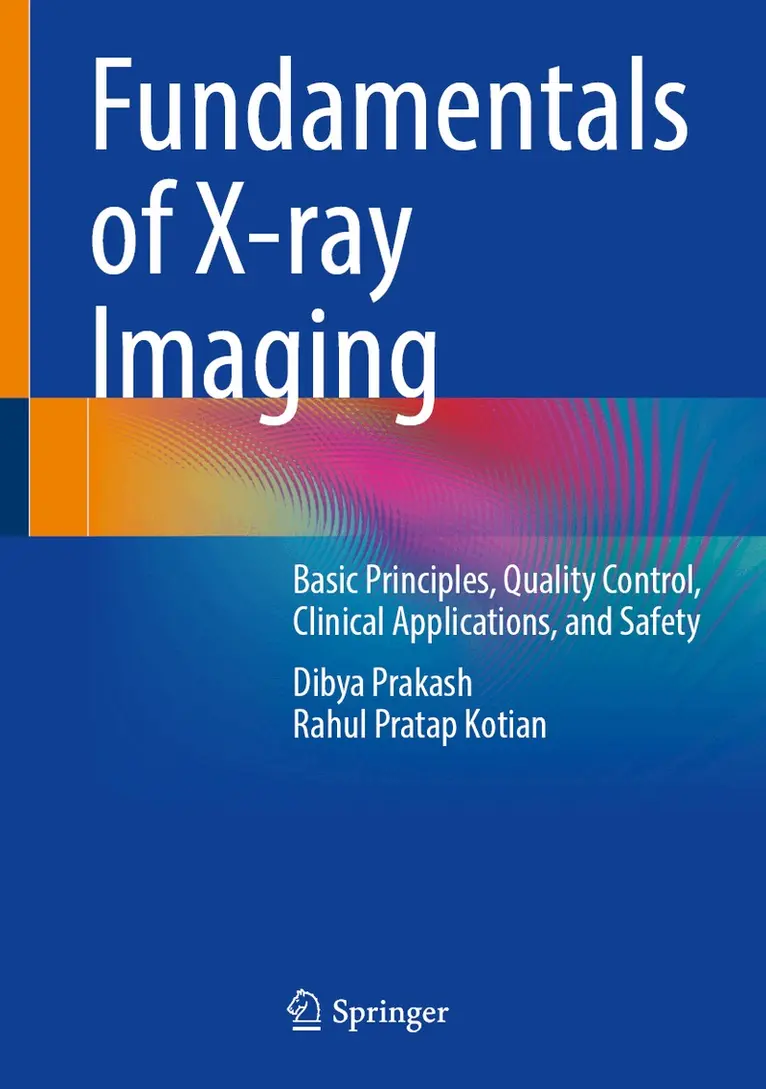
Fundamentals of X-ray Imaging
Basic Principles, Quality Control, Clinical Applications, and Safety
Av Dibya Prakash, Rahul Pratap Kotian
1336 kr
Beställningsvara. Skickas inom 10-15 vardagar. Fri frakt över 249 kr.
Beskrivning
Produktinformation
- Utgivningsdatum: 2025-09-27
- Mått: 178 x 254 x 42 mm
- Vikt: 1 619 g
- Format: Inbunden
- Språk: Engelska
- Antal sidor: 686
- Förlag: Springer Nature Switzerland AG
- ISBN: 9789819673278
Utforska kategorier
Mer om författaren
Dibya Prakash is a Scientific Officer (Nuclear Medicine) and Radiological Safety Officer – High Dose Therapy at ACTREC, Tata Memorial Centre, Mumbai. He holds Master’s degree in Physics and Bioinformatics and a Diploma in Medical Radioisotope Technology from BARC, Mumbai. A distinguished member of the Indian College of Nuclear Medicine (MICNM), he served in the Indian Navy for 15.5 years as paramedical staff.He has authored several books, including Radiation Safety Guide for Nuclear Medicine Professionals, Nuclear Medicine: A Guide for Healthcare Professionals and Patients, Radiation Emergency Preparedness Plan for Nuclear Medicine, and Nuclear Medicine CT Physics. His contributions also include scientific papers and abstracts in peer-reviewed journals.As the founder of Nuclear Medicine Solutions (NM Solutions) since 2019, he developed online courses, including a Nuclear Medicine Dose Assessment Course with ten modules on internal dosimetry. He has created educational YouTube tutorials for patients and professionals worldwide, promoting knowledge sharing in nuclear medicine.Dibya Prakash is a life member of the Society of Nuclear Medicine, India, and an affiliate member of the World Federation of Nuclear Medicine and Biology and the Nuclear Medicine Physicist Association of India. He serves as faculty in the SMG for Medical Physics and a committee member of the Council of Nuclear Medicine Science and Technology, India.Dr. Rahul P. Kotian is a renowned expert in Medical Imaging Technology, serving as Customer Lifetime Success Manager – Imaging at Philips (Middle East, Turkey, and Africa), Dubai, UAE. He is also a Visiting Professor of Radiology and Imaging at several Indian universities. With over 15 years of experience, his research focuses on MRI and CT imaging, particularly for early Parkinson’s disease diagnosisDr. Rahul holds a PhD in Magnetic Resonance Imaging from Manipal College of Health Professions, Manipal Academy of Higher Education, India. His groundbreaking work on Fractional Anisotropy – Diffusion Tensor Imaging of the brain laid the foundation for his book Diffusion Tensor Imaging and Fractional Anisotropy: Imaging Biomarkers in Early Parkinson’s Disease (Springer Nature). He has published extensively in MRI, CT, Fluoroscopy, Mammography, Radiation Protection, and Nuclear Medicine.His research aims to improve early Parkinson’s detection through imaging protocols that enhance diagnostic accuracy. An active member of SCMR, RSNA, ECR, ARM, and Arab Health, he frequently presents at international conferences. He also mentors graduates, postgraduates, and doctoral students, guiding the next generation of medical imaging professionals. Outside work, Dr. Rahul enjoys cricket, fitness, and global travel, drawing inspiration from nature.
Betyg & recensioner
0 recensioner
Inga recensioner tillgängliga.