• Fri frakt över 249 kr
  • •
  • Snabba leveranser
  • •
  • Billiga böcker
Kundservice

Du är på sajten för privatpersoner.

Företag, bibliotek eller offentlig verksamhet?

Du handlar på classic.bokus.com, där alla dina funktioner finns intakta.
Till classic.bokus.com
Bokus logotyp. Gå till startsidan.
  • Erbjudanden
  • Nyheter
  • Student
  • Topplistor
  • Barn & ungdom
  • Bokus Play
  • E-böcker
  • Pocketböcker
  • Spel & pussel

10% rabatt på allt med kod: NYSTART10 →

Sidfot

Mina sidor

    Hjälp

    • Kundservice
    • Vanliga frågor och svar
    • Frakt och leverans
    • Retur vid ångerrätt
    • Reklamera vara
    • Betalning
    • Köpvillkor
    • Allmänna villkor
    • Information om webbplatsens tillgänglighet

    Om Bokus

    • Om oss
    • Pressrum
    • För studenter
    • För företag
    • För bibliotek och offentlig verksamhet
    • För leverantörer
    • Hållbarhet

    Populärt

    • Aktuella erbjudanden
    • Presentkort
    • Studentlitteratur
    • Nya böcker
    • Topplistor
    • Signerade böcker
    • Engelska böcker

    Inspiration

    • Boktips
    • BookTok
    • Populära bokserier
    • Barnbokskaraktärer
    • Populära författare
    Logotyp för Bokus
    Följ oss på Facebook (extern länk)Följ oss på Instagram (extern länk)Följ oss på YouTube (extern länk)Följ oss på TikTok (extern länk)
    bokus @ CookiesAnpassa cookiesIntegritetspolicyKöpvillkor
    Till Citymail hemsida (extern länk)Till Budbee hemsida (extern länk)Till Postnord hemsida (extern länk)Till Schenker hemsida (extern länk)Till Early Bird hemsida (extern länk)Till Walleys hemsida (extern länk)
    1. Medicin
    2. Andra medicinska specialiteter
    3. Nuklearmedicin

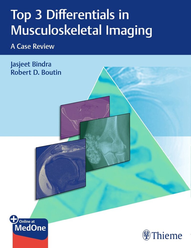
    Top 3 Differentials in Musculoskeletal Imaging

    A Case Review

    AvJasjeet Bindra,Robert D. Boutin

    Thieme Medical Publishers Inc

    2021

    Del i serien Top 3 Differentials

    1 188 kr

    Beställningsvara. Skickas inom 5-8 vardagar. Fri frakt över 249 kr.

    Beskrivning

    The highest-yield musculoskeletal radiology exam prep and learning tool available today!Top 3 Differentials in Musculoskeletal Imaging: A Case Review by Jasjeet Bindra, Robert D. Boutin, and expert contributors is one in a series of radiology case books mirroring the format of the highly acclaimed O'Brien classic, Top 3 Differentials in Radiology: A Case Review. The book is organized in 10 parts: trauma, bone tumors, upper extremity, lower extremity, arthropathies, infection, soft tissue tumors, metabolic musculoskeletal conditions, spine, and pediatric/developmental musculoskeletal conditions.Each case is formatted as a two-page unit. The left page features clinical images, succinctly captioned findings, and pertinent clinical history. The right page includes the key imaging gamut, differential diagnoses, additional diagnostic considerations, the diagnosis, clinical pearls, and suggested readings.Key FeaturesA total of 146 cases, each focused on a specific imaging finding, including aggressive periosteal reaction, focal cortical thickening, diffusely increased bone density, focal periphyseal edema, acro-osteolysis, and moreA wealth of meticulously selected, high-quality radiographs, CTs, and MRIs enhance diagnostic skillsA list of differential diagnoses provides an ideal curriculum guide for trainees and educators alikeRadiology residents, fellows, and staff radiologists preparing for certification will greatly benefit from reading this text to prep for the radiology core and certifying exams. This is also an outstanding, day-to-day practice resource for practicing radiologists, clinicians, and orthopaedic surgeons involved in reviewing and interpreting musculoskeletal radiology studies.This book includes complimentary access to a digital copy on https://medone.thieme.com.

    Produktinformation

    • Märke:Thieme Medical Publishers Inc
    • Utgivningsdatum:2021-02-24
    • Höjd:213 x 277 x 18 mm
    • Vikt:1 022 g
    • Språk:Engelska
    • Serie:Top 3 Differentials
    • Antal sidor:330
    • Förlag:Thieme Medical Publishers Inc
    • EAN:9781626233485

    Utforska kategorier

    • Nuklearmedicin inom Medicin
    • Reumatologi inom Medicin
    • Medicinsk bildbehandling inom Medicin

    Innehållsförteckning

    • Part 1: Trauma1 Shoulder dislocation2 Distal clavicular erosion3 Widening of acromioclavicular (AC) joint4 Fracture dislocation of forearm5 Carpal dislocation6 Avulsion fracture of finger7 Avulsion fracture of the pelvis8 Widened pubic symphysis9 Pelvic ring disruption10 Avulsion fracture at the knee11 Distal fibular fracture12 Calcaneal avulsion fracture13 Fracture at the base of the fifth metatarsalPart 2: Bone Tumors14 Aggressive periosteal reaction15 Focal cortical thickening16 Epiphyseal equivalent lucent lesion17 Solitary focal sclerotic lesion18 Lesion with permeative or moth-eaten pattern19 Osseous lesion with fluid–fluid levels20 Osseous lesion with chondroid matrix21 Osseous lesion with osteoid matrix22 Polyostotic lesions–looks like a syndrome23 Surface lesion of bone24 Multiple lytic lesions25 Multiple sclerotic lesions26 Expansile lytic lesion of rib27 Ivory vertebral body28 Lytic lesion of distal phalanx29 Anterior tibial cystic appearing lesion30 Lucent lesion in calcaneus31 Diffusely increased bone density32 Well-marginated ossific mass projecting off the surface of the bone (without full-thickness cortical disruption or overlying soft-tissue mass)33 Long lesion in a long bone with ground glass matrix–Roentgen classic34 Bony projection with continuation of cortex and medullary cavity of host bone–Roentgen classic35 Fractured lytic lesion with a fallen fragment–Roentgen classicPart 3: Upper Extremity36 Abnormal/variant appearance of anterosuperior labrum37 Abnormality of inferior glenohumeral ligament38 Abnormality of proximal long head of biceps tendon39 Tear of the anteroinferior labrum40 Bursa or cyst around the shoulder41 Rotator cuff abnormality42 Rotator interval abnormality43 Posterior glenoid labral abnormality44 Nerve entrapment at shoulder45 Nerve entrapment at elbow46 Distal biceps tendon abnormality47 Radial-sided, soft-tissue injury at elbow on MRI48 Osteochondral lesion of elbow49 Ulnar-sided, stabilizer injury at elbow on MRI50 Numerous tiny T2 hyperintense subcutaneous nodulesPart 4: Lower Extremity51 Variant sciatic nerve anatomy52 Ischiofemoral space narrowing and quadratus femoris muscle edema in a patient with chronic ischial tuberosity avulsion (nonunion)53 Small smoothly marginated depression ("fossa") at the mid-acetabular roof54 Focal subchondral bone edema in the femoral head55 Low-grade tearing of the iliotibial band (ITB), with soft-tissue edema along the fascia56 Focal periphyseal edema57 Curvilinear signal separating the patella into two fragments58 Frond-like lobules of fatty tissue in the suprapatellar recess, with effusion59 Cystic-appearing lesion centered inferior to the medial joint line of the knee60 Accessory muscle around ankle61 Accessory ossicle around ankle62 Achilles tendon abnormality63 Impingement syndrome at ankle64 Ankle sprain65 Medial tendon abnormality at ankle66 Nerve entrapment around ankle and foot67 Plantar fascial abnormality68 Peroneal tendon disease at the ankle69 Midfoot ligamentous or osseous injury70 Hallux sesamoid abnormality71 Subchondral crescent sign–Roentgen classic72 Fluid collection at the inferomedial aspect of hip–Roentgen classic73 Cleft of T2 hyperintense signal at the attachment of rectus abdominis-adductor aponeurosis–Roentgen classic74 Hamstring tendon discontinuity with subacute hematoma–Roentgen classicPart 5: Arthropathies75 Intense, diffuse, nontraumatic bone and soft-tissue edema centered at the midfoot76 Polyarticular arthritis in an adolescent, with "blooming" intraarticular hemosiderin77 Bilateral sacroiliitis78 Contour defect at the posterolateral humeral head79 Proximal arthropathy affecting primarily the metacarpophalangeal joints.80 Distal arthropathy affecting primarily the interphalangeal joints81 Erosive arthropathy involving the foot82 Acro-osteolysis83 Loose bodies84 Articular cartilage thinning, subchondral cyst formation and dorsal subluxation of the metacarpal base–Roentgen classicPart 6: Infection85 Penumbra sign86 Sequestrum87 Fluid collection with edema at metaphyseal-equivalent location (prior to skeletal maturity)88 Space occupying lesion posterior to olecranon89 Unilateral sacroiliitis90 Epitrochlear lymphadenopathy91 Aggressive, cortically-based osteolysis and periosteal reaction at the diaphysis of an adult long bone92 Abscesses in bone and adjacent to bone, with thin perforations in cortex–Roentgen classicPart 7: Soft Tissue Tumors93 Fusiform mass, with longitudinal "tail" entering and exiting eccentrically94 Complex fluid collection in the bicipitoradial bursa95 Infiltrative intra-articular mass of low-T2 signal intensity, with erosions96 Non-mineralized soft-tissue mass with an infiltrative border ("tail sign") that responds favorably to non-operative treatment97 Post-traumatic fluid-like collection in the deep subcutaneous tissue along the superficial aspect of the fascia98 Large, pendulous mass at the inner thigh in a morbidly obese patient99 Leg, ankle, and foot edema, with prominent thickening and increased density of the subcutaneous fat layer100 Triad of key findings: Pretibial varices, linear cortical defect in mid tibia, and prominent nutrient vein within the mid tibia101 Multiple T2-hypointense masses in the subcutaneous fat layer of the foot102 Large subfascial soft-tissue mass with heterogeneous signal on T1, T2, and post-contrast images103 Rim enhancing mass in iliopsoas104 Infrascapular soft-tissue mass containing diffuse streaks of fatty tissue–Roentgen classicPart 8: Metabolic Musculoskeletal Conditions105 Frayed metaphyses106 Generalized periosteal reaction107 Periarticular calcified mass108 Long bone sclerosis109 Multiple bone infarcts110 Osteoporosis111 Widespread soft tissue calcifications112 Rugger jersey spinePart 9: Spine113 Vertebral body fusion114 Odontoid fracture115 Hyperflexion Injury116 Midline sacral lesion117 Posterior vertebral body scalloping118 Anterior vertebral body scalloping119 Lytic lesion of posterior elements of spine120 Epidural mass121 Diffuse marrow T1 and T2 low signal122 Focal T1 signal increase123 Intradural extramedullary mass124 Intradural intramedullary spinal cord mass125 End plate irregularityPart 10: Pediatric or Developmental Musculoskeletal Conditions126 Short rib skeletal dysplasia127 Abnormal epiphyses128 Limb deficiency129 Genu varum130 Lucent metaphyses131 Dense metaphyses132 Dislocated hip133 Physeal widening134 Hip effusion135 Infrapatellar soft tissue swelling136 Foot deformity137 Soft tissue mass in an infant138 Large soft tissue mass in an older child139 Periosteal reaction in a newborn/infant140 Wormian bones141 Madelung deformity142 Tarsal coalition143 Well-corticated ossific density at the lateral aspect of acromion–Roentgen classic144 Osteoporosis, bowing deformities, and multilevel spine fractures–Roentgen classic145 Decreased lower lumbar interpediculate distances, "tombstone" iliac bones, "champagne glass" pelvic inlet, and rhizomelic limb shortening–Roentgen classic146 Multiple rib fractures in various stages of healing–Roentgen classic