• Fri frakt över 249 kr
  • •
  • Snabba leveranser
  • •
  • Billiga böcker
Kundservice

Du är på sajten för privatpersoner.

Företag, bibliotek eller offentlig verksamhet?

Du handlar på classic.bokus.com, där alla dina funktioner finns intakta.
Till classic.bokus.com
Bokus logotyp. Gå till startsidan.
  • Erbjudanden
  • Nyheter
  • Student
  • Topplistor
  • Barn & ungdom
  • Bokus Play
  • E-böcker
  • Pocketböcker
  • Spel & pussel

10% studentrabatt med kod TERM26

Sidfot

Mina sidor

    Hjälp

    • Kundservice
    • Vanliga frågor och svar
    • Frakt och leverans
    • Retur vid ångerrätt
    • Reklamera vara
    • Betalning
    • Köpvillkor
    • Allmänna villkor
    • Information om webbplatsens tillgänglighet

    Om Bokus

    • Om oss
    • Pressrum
    • För studenter
    • För företag
    • För bibliotek och offentlig verksamhet
    • För leverantörer
    • Hållbarhet

    Populärt

    • Aktuella erbjudanden
    • Presentkort
    • Studentlitteratur
    • Nya böcker
    • Topplistor
    • Signerade böcker
    • Engelska böcker

    Inspiration

    • Boktips
    • BookTok
    • Populära bokserier
    • Barnbokskaraktärer
    • Populära författare
    Logotyp för Bokus
    Följ oss på Facebook (extern länk)Följ oss på Instagram (extern länk)Följ oss på YouTube (extern länk)Följ oss på TikTok (extern länk)
    bokus @ CookiesAnpassa cookiesIntegritetspolicyKöpvillkor
    Till Citymail hemsida (extern länk)Till Budbee hemsida (extern länk)Till Postnord hemsida (extern länk)Till Schenker hemsida (extern länk)Till Early Bird hemsida (extern länk)Till Walleys hemsida (extern länk)
    1. Medicin
    2. Veterinärmedicin
    3. Veterinärmedicin: smådjur

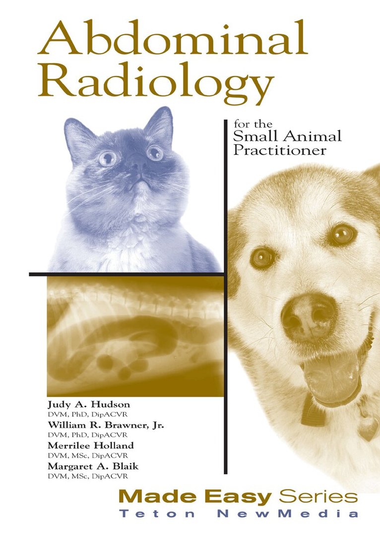
    Abdominal Radiology for the Small Animal Practitioner

    AvJudith Hudson,William Brawner

    Teton NewMedia

    2001

    Del i serien Made Easy Series

    1 080 kr

    Tillfälligt slut

    Fler format och utgåvor

    Häftad

    Tillf. slut

    Beskrivning

    This text is meant to be a handy cookbook which can be quickly grabbed from the self and provide the practitioner with essential information on abdominal radiography. This practical presentation consists of the uses and interpretations of abdominal plain film for the small animal practitioner or technician. The text describes the normal appearance of the abdomen, ways in which the radiographic appearance changes to reflect disease, and common abdominal disorders. Discussions in the text also include techniques for better radiographs and steps to good film reading. The lay-flat binding is ideal for practical use in the clinic.Published by Teton New Media in the USA and distributed by CRC Press outside of North America.

    Produktinformation

    • Märke:Teton NewMedia
    • Utgivningsdatum:2001-12-11
    • Höjd:153 x 216 x 10 mm
    • Vikt:363 g
    • Språk:Engelska
    • Serie:Made Easy Series
    • Antal sidor:157
    • Förlag:Teton NewMedia
    • EAN:9781893441347

    Utforska kategorier

    • Veterinärmedicin: smådjur inom Medicin
    • Veterinärmedicin inom Medicin

    Innehållsförteckning

    • Section1 Introduction andRadiographic TechniqueSome Helpful HintsIndications for Abdominal RadiographyRole of Radiology in Patient ManagementSteps to Good Film ReadingStep One: Technical Factors forAbdominal RadiographyStep Two: Using a System for InterpretationStep Three: Roentgen or Radiographic SignsStep Four: Differential DiagnosesStep Five: What’s Next?Section2Normal RadiographicAnatomy of the AbdomenViewing the FilmStomachDuodenumCecumKidneySpleenDiaphragmLiverBladderProstateLymph NodesSection3Peritoneal CavityNormal AppearanceIncreased Peritoneal OpacityTerminology – SynonymsDecreased Peritoneal Opacity–GasCauses of Intraluminal Gas AccumulationCauses of Extraluminal Gas AccumulationRadiographic (Roentgen)Signs of Extraluminal GasDecreased Peritoneal Opacity–FatCauses of Abnormal Fat OpacitiesRadiographic (Roentgen) SignsDisruption of Borders of the Peritoneal CavityDiaphragmatic HerniaHiatal HerniaPeritoneopericardial HerniaInguinal or Ventral HerniaPerineal HerniaSection4 Intra-abdominal MassesEvaluation of an Abdominal MassGastric MassesGeneralized HepatomegalyFocal HepatomegalyDifferentiate the StomachRenal MassesAdrenal MassDiffuse SplenomegalyFocal SplenomegalyMesenteric/Enteric MassesPancreatic MassesOvarian MassesMasses Involving Urinary BladderProstatic MassesUterine MassesCaudal Sublumbar MassesSection5Alimentary TractContrast MediaBariumIonic Organic IodineNon-Ionic Organic Iodine PreparationsEsophageal/Gastrointestinal Contrast ProceduresRadiography of the EsophagusContrast Examination of the Esophagus–EsophagramEsophagram TechniqueDisorders of the EsophagusEsophageal Foreign BodiesMegaesophagusVascular Ring AnomaliesEsophageal MassesRadiography of the Stomach and Small IntestineSurvey RadiographsContrast Examination of theStomach and Small Intestine–IndicationsUpper Gastrointestinal SeriesNormal Upper GI SeriesStomachSmall IntestineDifferences in CatsPrinciples of InterpretationOther Contrast ProceduresUpper GI Series with IodinePneumogastrogramDouble Contrast GastrogramDisorders of the StomachGastric Foreign BodyGastric Torsion/DilatationPyloric Outflow ObstructionGastric NeoplasiaGastroesophageal IntussusceptionDisorders of Small IntestineIleusMechanical ObstructionForeign BodyIntussusceptionInflammatory Diseases Without UlcerationUlcersInfiltrative DiseaseRadiography of the Large IntestineContrast Radiography of the Large IntestinePneumocolographyBarium EnemaDouble Contrast Barium EnemaDisorders of the Large IntestineObstipationIleocolic IntussusceptionCecal InversionInfiltrative DiseasesMucosal Diseases (Colitis)Section6Urinary TractSelection of Appropriate Contrast ProcedureContrast Examination of the Urinary Bladder(Cystography) Positive Contrast CystogramNegative Contrast Cystogram (Pneumocystogram)Double Contrast CystogramVesicoureteral RefluxDisorders of the Urinary BladderUrinary CalculiRuptured BladderCystitisEmphysematous CystitisUrinary Bladder NeoplasiaContrast Examination of the Urethra (Urethrography)Disorders of the UrethraUrethral CalculiObstructive UropathyRuptured UrethraContrast Examination of the Kidneys andUreters–Excretory UrogramNormal Excretory UrogramArteriogram PhaseNephrogram PhasePyelogram PhaseCystogram PhaseDisorders of the Kidneys and UretersChronic Renal DiseaseRenal DysplasiaTransitional Cell Carcinoma of the Urinary BladderHydronephrosisRenal CalculiPyelonephritisRenal NeoplasiaPolycystic Renal DiseasePerirenal PseudocystCompensatory HypertrophyFunctional Evaluation of the KidneyRuptured UreterUreteral IleusUreteral CalculiPrimary UreteritisEctopic UreterSection7Reproductive TractFemale: Uterus and OvariesPregnancyDisorders of the Female Reproductive TractPyometra and Other Causes ofUterine EnlargementDystociaFetal DeathOvarian NeoplasiaDisorders of the Male Reproductive TractRetained Testicles and Prostate GlandTesticular MassesProstatic EnlargementBenign Prostatic HyperplasiaProstatitisProstatic AbscessProstatic NeoplasiaProstatic and Paraprostatic CystsSection8AnomaliesMicrohepaticaRenal AgenesisMalpositioned KidneysKartagener’s SyndromeRecommended Readings