E-bok, Engelska, 2022
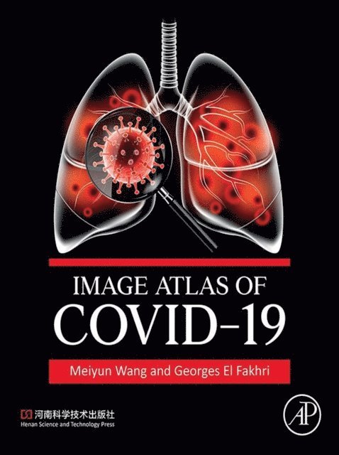
Image Atlas of COVID-19
Av Georges El Fakhri, Meiyun Wang
2402 kr
Läs direkt i Bokus Reader – eller ladda ned till din enhet
Beskrivning
Image Atlas of COVID-19 covers this novel and emerging respiratory infectious disease. Although the gold standard for clinical diagnosis of COVID-19 remains the nucleic acid test for SARS-CoV-2, chest CT scans have been widely used in the diagnosis and treatment of COVID-19 as simple and fast diagnostic methods. The latter has proven crucial when faced with a large volume of symptomatic and asymptomatic patients. The imaging manifestations of COVID-19 have some specific characteristics, hence a better understanding of imaging findings is of great value for detecting lesions, evaluating lesion severity, diagnosis and differential diagnosis, and assessment of therapeutic options and prognosis.- Contains 61 confirmed cases of COVID-19, including a collection of over 400 DR, CT, PET/CT and PET/MRI scans in Henan Provincial People''s Hospital (HPPH)- Organized into four chapters by clinical subtypes of COVID-19- Details a large number of clinical images, with illustrations that are intended to demonstrate imaging findings of COVID-19 to serve as a reference manual for medical staff engaged in imaging and clinical work- Ideal as a training material for academic education
Produktinformation
- Utgivningsdatum: 2022-09-21
- Språk: Engelska
- Filformat: EPUB
- Kopieringsskydd: LCP
- ISBN: 9780443184925
- Förlag: Elsevier Science
Utforska kategorier
Betyg & recensioner
0 recensioner
Inga recensioner tillgängliga.