Häftad, Engelska, 2000
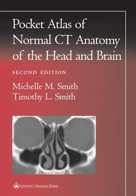
Pocket Atlas of Normal CT Anatomy of the Head and Brain
Av Michelle M. Smith, Timothy L. Smith
457 kr
Beställningsvara. Skickas inom 7-10 vardagar. Fri frakt över 249 kr.
Beskrivning
Featuring 73 sharp, new images obtained with state-of-the-art scanning technology, the Second Edition of this popular pocket atlas is a quick, handy guide to interpreting computed tomography images of the brain and calvarium, temporal bone, orbit, nasal cavity, and paranasal sinuses. The book helps readers recognize normal anatomic structures on CT scans and distinguish these structures from artifacts.Each page presents a high-resolution CT scan, with anatomic landmarks clearly labeled. Directly above the scan are a key to the labels and a thumbnail illustration that orients the reader to the plane of view (sagittal, axial, or coronal). This format enables readers to identify features rapidly and accurately. Praise for the previous edition: "[A] splendid, very pocket-sized atlas....[A] very useful basic booklet which will enlighten all clinical trainees and not a few of their mentors."-- Journal of Neurology, Neurosurgery, and Psychiatry "A very useful and uncomplicated pocket-sized reference....This little atlas will be popular among residents in neurology, neurosurgery, ENT, ophthalmology, and internal medicine, and all clinicians who are required to inspect CTs of the head."-- Neurology "The book is most useful for medical students and neophyte radiology, neurology, or neurosurgical residents....Recommended!-- American Journal of Neuroradiology
Produktinformation
- Utgivningsdatum: 2000-09-23
- Mått: 124 x 175 x 5 mm
- Vikt: 91 g
- Format: Häftad
- Språk: Engelska
- Serie: Radiology Pocket Atlas Series
- Antal sidor: 88
- Upplaga: 2
- Förlag: Lippincott Williams and Wilkins
- ISBN: 9780781729499
Utforska kategorier
Betyg & recensioner
0 recensioner
Inga recensioner tillgängliga.