Häftad, Engelska, 2016
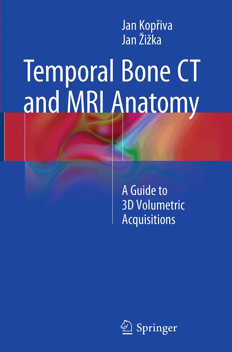
Temporal Bone CT and MRI Anatomy
Av Jan Kopřiva, Jan Žižka
917 kr
Skickas inom 10-15 vardagar
Beskrivning
This book, featuring more than 180 high spatial resolution images obtained with state-of-the-art MDCT and MRI scanners, depicts in superb detail the anatomy of the temporal bone, recognized to be one of the most complex anatomic areas. In order to facilitate identification of individual anatomic structures, the images are presented in the same way in which they emanate from contemporary imaging modalities, namely as consecutive submillimeter sections in standardized slice orientations, with all anatomic landmarks labeled. While various previous publications have addressed the topic of temporal bone anatomy, none has presented complete isotropic submillimeter 3D volume datasets of MDCT or MRI examinations. The Temporal Bone MDCT and MRI Anatomy offers radiologists, head and neck surgeons, neurosurgeons, and anatomists a comprehensive guide to temporal bone sectional anatomy that resembles as closely as possible the way in which it is now routinely reviewed, i.e., on the screens of diagnostic workstations or picture archiving and communication systems (PACS).
Produktinformation
- Utgivningsdatum: 2016-08-23
- Mått: 155 x 235 x 13 mm
- Vikt: 353 g
- Format: Häftad
- Språk: Engelska
- Antal sidor: 203
- Förlag: Springer International Publishing AG
- ISBN: 9783319361604
Utforska kategorier
Betyg & recensioner
0 recensioner
Inga recensioner tillgängliga.