• Fri frakt över 249 kr
  • •
  • Snabba leveranser
  • •
  • Billiga böcker
Kundservice

Du är på sajten för privatpersoner.

Företag, bibliotek eller offentlig verksamhet?

Du handlar på classic.bokus.com, där alla dina funktioner finns intakta.
Till classic.bokus.com
Bokus logotyp. Gå till startsidan.
  • Erbjudanden
  • Nyheter
  • Student
  • Topplistor
  • Barn & ungdom
  • Bokus Play
  • E-böcker
  • Pocketböcker
  • Spel & pussel

Må bättre, för mindre! Upp till 50% rabatt på hälsoböcker

Sidfot

Mina sidor

    Hjälp

    • Kundservice
    • Vanliga frågor och svar
    • Frakt och leverans
    • Retur vid ångerrätt
    • Reklamera vara
    • Betalning
    • Köpvillkor
    • Allmänna villkor
    • Information om webbplatsens tillgänglighet

    Om Bokus

    • Om oss
    • Pressrum
    • För studenter
    • För företag
    • För bibliotek och offentlig verksamhet
    • För leverantörer
    • Hållbarhet

    Populärt

    • Aktuella erbjudanden
    • Presentkort
    • Studentlitteratur
    • Nya böcker
    • Topplistor
    • Signerade böcker
    • Engelska böcker

    Inspiration

    • Boktips
    • BookTok
    • Populära bokserier
    • Barnbokskaraktärer
    • Populära författare
    Logotyp för Bokus
    Följ oss på Facebook (extern länk)Följ oss på Instagram (extern länk)Följ oss på YouTube (extern länk)Följ oss på TikTok (extern länk)
    bokus @ CookiesAnpassa cookiesIntegritetspolicyKöpvillkor
    Till Citymail hemsida (extern länk)Till Budbee hemsida (extern länk)Till Postnord hemsida (extern länk)Till Schenker hemsida (extern länk)Till Early Bird hemsida (extern länk)Till Walleys hemsida (extern länk)
    1. Medicin
    2. Kirurgi
    3. Gastrointestinal och kolorektal kirurgi

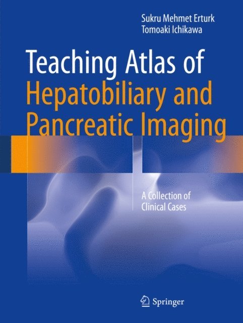
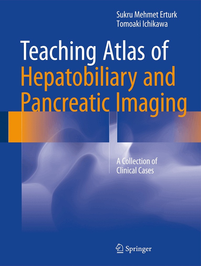
    Teaching Atlas of Hepatobiliary and Pancreatic Imaging

    A Collection of Clinical Cases

    AvSukru Mehmet Erturk,Tomoaki Ichikawa

    Häftad, Engelska, 2018

    1 007 kr

    Beställningsvara. Skickas inom 10-15 vardagar. Fri frakt över 249 kr.

    Fler format och utgåvor

    Inbunden

    1 452 kr

    Beskrivning

    Featuring 137 carefully selected cases, this atlas covers virtually every aspect of clinical cross-sectional imaging of the liver, gallbladder, biliary system and pancreas. For the vast majority of the cases, both CT and MR images are included to demonstrate the different features of each lesion. Furthermore, both typical and atypical pathologies are included to facilitate the differential diagnosis in daily clinical practice. Concise yet comprehensive,this atlas includes not only imaging features of the lesions but also the related pathologic and clinical data. It is therefore useful both as a quick guide for practicing radiologists and as a brief textbook for radiologists in training.

    Produktinformation

    • Utgivningsdatum:2018-06-12
    • Mått:210 x 279 x 12 mm
    • Vikt:645 g
    • Format:Häftad
    • Språk:Engelska
    • Antal sidor:223
    • Förlag:Springer International Publishing AG
    • ISBN:9783319820156

    Utforska kategorier

    • Gastrointestinal och kolorektal kirurgi inom Medicin
    • Allmänkirurgi inom Medicin
    • Klinisk medicin och internmedicin inom Medicin

    Mer om författaren

    Sukru Mehmet Erturk studied medicine at Cerrahpasa School of Medicine, Istanbul University, and then completed a one-year certificate program in hospital management at the Institute of Business Administration of the same university. Mehmet started his specialty training in radiology at the Sisli Hamidiye Etfal Training and Research Hospital in 1998. In 2001, he was an observer at the Radiology Department of the Klinikum Grosshadern of Ludwig Maximillian University of Munich. Upon completing his training and his work as an attending radiologist at Sisli Hamidiye Etfal Training and Research Hospital, Mehmet moved to Boston and completed his fellowships in management in radiology and in abdominal radiology research between 2004 and 2006 at the Brigham and Women’s Hospital and the Harvard Medical School, where he was also a lecturer in radiology.  In 2007, he was elected as president of the Istanbul Chapter of the Turkish Society of Radiology and in 2009 he became the Secretary Generalof the same society, of which he is still an executive board member. Mehmet’s areas of research and interest include topics such as abdominal imaging, utilization management in radiology and health services, quality management in radiology and health services, medical education, health economics, and health informatics. He holds editorial and reviewer positions in various journals. Currently, he is Professor of Radiology and Chairman of Radiology Department at Adiyaman University, Turkey.Tomoaki Ichikawa is Associate Professor and Chief of the Diagnostic Radiology Division, Department of Radiology, University of Yamanashi, Yamanashi, Japan. His principal area of interest is hepatobiliary CT/MR imaging. Dr. Ichikawa completed his medical studies at the Graduate School of Medicine and School of Medicine, Chiba University, Japan in 1988 and subsequently specialized in Radiology. In 1998 he became a Research Fellow in the Division of Abdominal Radiology, Department ofRadiology,University of Pittsburgh Medical Center, Pittsburgh, USA. He was subsequently appointed Assistant Professor and then Associate Professor and Head of the Abdominal Imaging Division in the Department of Radiology at University of Yamanashi. In 2004 he became Visiting Associate Professor in the Department of Radiology, Brigham & Women's Hospital, Harvard Medical School. He took up his present post in 2006. Dr. Ichikawa is an editorial board member and reviewer for the journal Investigative Radiology. He has published approximately 90 publications in peer-reviewed English language journals.

    Innehållsförteckning

    • Part I Liver: Simple cyst of the liver.- Polycystic liver disease.- Hemangioma 1.- Hemangioma 2.- Hemangioma 3.- Hemangioma 4.- FNH 1.- FNH 2.- FNH 3.- Hepatic adenoma 1.- Hepatic adenoma 2.- Hepatic lipoma.- Sarcoidosis.- Infantile hemangioendothelioma.- Diffuse hepatosteatosis.- Budd-Chiari.- Venoocclusive disease liver.- Wilson disease.- Focal steatosis.- Focal fatty sparing.- Secondary hemochromatosis.- Vascular injury.- Cirrhosis .- Cirrhosis (advanced).- Cavernous transformation of the portal vein.- Dysplastic nodule.- Confluent hepatic fibrosis.- HCC 1.- HCC 2 (well differentiated).- HCC 3 (diffuse).- Fibrolamellar HCC.- Hepatoblastoma.- Hepatic lymphoma.- Primary neuroendocrine tumor of the liver.- Liver metastasis (hypovascular).- Liver metastasis (hypervascular).- Liver metastasis (neuroendocrine tumor).- Pseudocirrhosis.- Cavernous transformation of the portal vein.- Pyogenic abscess of the liver.- Amebic abscess of the liver.- Hepatic candidiasis.- Schistosomiasis.- Fascioliasis.- Hydatid cyst 1.- Hydatid cyst 2.- Hydatid cyst 3.- Hydatid cyst 4.- Echinococcus alveolaris.- Part II Gallbladder and biliary system:  Acute cholecystitis and cholelithiasis.- Gangrenous cholecystitis.- Emphysematous cholecystitis.- Acute cholecystitis and perforation of gall bladder.- Acute cholecystitis and intrahepatic abscess formation.- Choledocholithiasis.- Stone in the ductus cysticus.- Mirizzi syndrome.- Xantagranulamatous cholecystisi.- Porcelain gallbladder.- Tumefactive sludge within the gallbladder.- Gallbladder polyp.- Gallbladder cancer 1.- Gallbladder cancer 2.- Gallbladder cancer with perforation.- Biliary cystadenoma-cystadenocarcinoma.- Intrahepatic cholangiocarcinoma 1.- Intrahepatic cholangiocarcinoma 2.- Perihiler cholangiocarcinoma Type 1.- Perihiler cholangiocarcinoma Type 2.- Perihiler cholangiocarcinoma Type 3a.- Perihiler cholangiocarcinoma Type 3b.- Perihiler cholangiocarcinoma Type 4.- Type 1 choledochal cyst.- Type 2 choledochal cyst.- Type 3 choledochal cyst.- Type 4 choledochal cyst.- Type 5 choledochal cyst (Caroli disease).- Caroli syndrome.- Common bile duct adenoma.- Von-Meyenburg complex.- Primary sclerosing cholangitis.- HIV-cholangiopathy.- Oriental cholangiohepatitis.- Part III Pancreas: Acute pancreatitis.- Hemorrhagic pancreatitis.- Necrotizing pancreatitis.- Pseudocyst 1.- Infected pseudocyst.- Groove pancreatitis.- Chronic pancreatitis 1.- Chronic pancreatitis 2.- Autoimmune pancreatitis.- Mucinous cystic neoplasm 1.- Mucinous cystic neoplasm 2.- Serous cystic neoplasm 1.- Serous cystic neoplasm 2.- IPMN: main duct type.- IPMN: side branch type.- IPMN: combined type.- Solid paillary epithelial neoplasm 1.- Solid papillary epithelial neoplasm 2.- Metastasis to pancreas 1.- Metastasis to pancreas 2.- Pancreas divisum.- Annular pancreas.- Pancreatic cysts in von Hippel Lindau disease.