Albert L. Rhoton – författare
3 453 kr
Skickas inom 5-8 vardagar
1 137 kr
Skickas inom 5-8 vardagar
3 895 kr
Läs direkt efter köp
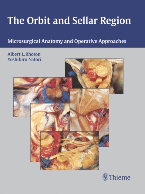
Designed to bring all of orbital anatomy into perspective, this expert reference is the first to:
1) Provide a comprehensive review of the microsurgical anatomy of the orbit and sellar region;2) Demonstrate the relationship of the orbit and surrounding structures; and3) Illustrate orbital structures from multiple operative approaches Hundreds of vivid dissections show the orbit from above, below, laterally, medially, and anteriorly, with illustrations fully labeled for valuable review and study.The organization of THE ORBIT AND SELLAR REGION leads to clarity and comprehension. Divided into three sections, the book begins with a full description of osseous, neural, arterial, venous, and muscular anatomy. It then goes on to stepwise dissections of the orbit from different directions, in which each layer is peeled away to expose the next deeper layer and the placement of the orbit and concludes with multiple common operative approaches to the sellar region.
1 645 kr
Läs direkt efter köp
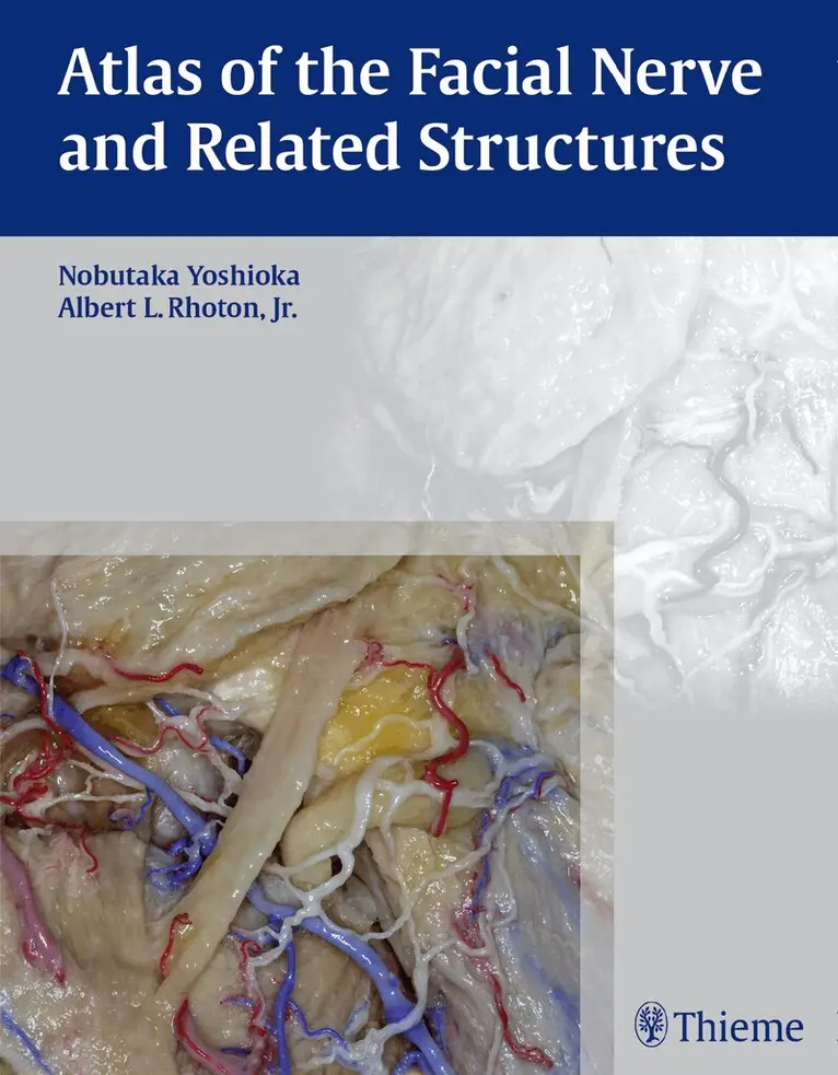
Nobutaka Yoshioka, MD, PhD and Albert L. Rhoton Jr., MD have created an anatomical atlas of astounding precision. An unparalleled teaching tool, this atlas opens a unique window into the anatomical intricacies of complex facial nerves and related structures.
An internationally renowned author, educator, brain anatomist, and neurosurgeon, Dr. Rhoton is regarded by colleagues as one of the fathers of modern microscopic neurosurgery. Dr. Yoshioka, an esteemed craniofacial reconstructive surgeon in Japan, mastered this precise dissection technique while undertaking a fellowship at Dr. Rhoton''s microanatomy lab, writing in the preface that within such precision images lies potential for surgical innovation.
Special Features:
Exquisite color photographs, prepared from carefully dissected latex injected cadavers, reveal anatomy layer by layer, with remarkable detail and clarity. An added highlight, 3-D versions of these extraordinary images are available online in the Thieme MediaCenter.Major sections include intracranial region and skull, upper facial and midfacial region, and lower facial and posterolateral neck region.Organized by region, each layered dissection elucidates specific nerves and structures with pinpoint accuracy, providing the clinician with in-depth anatomical insights. Precise clinical explanations accompany each photograph. In tandem, the images and text provide an excellent foundation for understanding the nerves and structures impacted by neurosurgical-related pathologies as well as other conditions and injuries.
An exceptionally stunning anatomical reference, this book is a must-have reference for residents, and advanced clinicians specializing in neurosurgery, facial plastic surgery, otolaryngology, maxillofacial surgery, and craniofacial surgery.
4 847 kr
Läs direkt efter köp
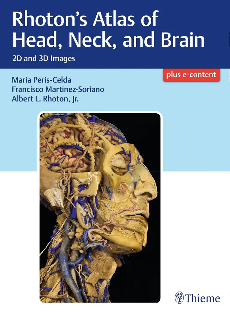
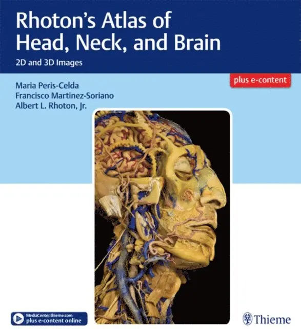
Masterful 2D and 3D head, neck, and brain dissections provide unsurpassed insights into head, neck, and brain anatomy
An internationally renowned and beloved author, educator, brain anatomist, and neurosurgeon, Professor Albert Rhoton has a special place in medical history. He was revered by students and colleagues and is regarded as one of the fathers of modern microscopic neurosurgery. A driving principle in his anatomy lab was the simple phrase, "Every Second." This was embraced in his philosophy that every second of every day, a patient''s life was improved by a surgeon assisted by the anatomic knowledge his lab helped elucidate and distribute.
Rhoton''s Atlas of Head, Neck, and Brain is the visually exquisite crowning achievement of Dr. Rhoton''s brilliant career and unwavering dedication to the intertwined pursuits of surgical anatomy and neurosurgery. The atlas reflects the unparalleled contributions Dr. Rhoton made to the contemporary understanding of neurosurgical anatomy. Dr. Peris-Celda, with the collaboration of an impressive cadre of international multidisciplinary experts, worked closely under Dr. Rhoton''s tutelage on this project. This book is the culmination of 5 years of work and experience gleaned from more than 40 years of surgical anatomy research and exquisite dissection techniques performed in Dr. Rhoton''s laboratory.
Special Features
Each anatomic dissection meticulously labeled with English and Latin descriptors for easy cross referencing with other resources.Multiple views of the most complex regions of the head, neck, and brain provide a deeper understanding of anatomy.More than 600 anatomical images systematically organized in four major sections: Osteology of the Head and Neck; Face and Neck; Ear, Nose, Pharynx, Larynx, and Orbit; and Neuroanatomy and Cranial Base.Superb 2D images presented in a large printed format to optimize the viewing experience.3D digital images fully realize the beauty of the dissections and enhance the learning process.Specimens injected with colored silicone provide better visualization of arteries and veins.Breathtakingly stunning, this atlas is certain to be a treasured reference for medical students, residents, and clinicians specializing in neurosurgery, facial plastic surgery, otolaryngology, maxillofacial surgery, and craniofacial surgery for many years to come.