
Francis S. Weill – författare
Visar alla böcker från författaren Francis S. Weill. Handla med fri frakt och snabb leverans.
9 produkter
9 produkter
Häftad, Tyska, 1990
582 kr
Skickas inom 10-15 vardagar
Dieses Buch bietet, kurzgefaßt und übersichtlich in 13 Kapital gegliedert, eine umfassende Einführung in die Anwendung der Computertomographie im Thorakal- und Abdominalbereich; die im Text gegebenen Anleitungen werden großzügig durch didaktisch hervorragendes Bildmaterial illustriert. Das Buch wendet sich nicht nur an Ärzte, die in der radiologischen Fortbildung stehen, sondern ist darüber hinaus auch für zuweisende Ärzte anderer Fachrichtungen (Allgemeinärzte, Internisten, Chirurgen) von ganz besonderem Interesse, da es knapp und kompetent über die Indikationen und diagnostischen Möglichkeiten der Computertomographie informiert.
E-bok
PDF, Engelska, 20121 459 kr
Läs direkt efter köp
For the new English edition, this highly popular book has been thoroughly revised and updated to include the enormous progress seen recently in the field of ultrasonography. After an extensive technical introduction the book covers the sonoanatomy and ultrasonic symptomatology of the diseases of the digestive system and the abdominal vessels. New chapters deal with portal hypertension, the intestine, including appendicitis, endoscopic ultrasonography of carcinomas and abdominal abnormalities arising from AIDS. In such a fast-moving medical field, this detailed, extensively illustrated book will help both the experienced clinician and those in training to consolidate their knowledge of ultrasound in daily practice.
Häftad, Engelska, 2011
1 125 kr
Skickas inom 10-15 vardagar
For the new English edition, this highly popular book has been thoroughly revised and updated to include the enormous progress seen recently in the field of ultrasonography. After an extensive technical introduction the book covers the sonoanatomy and ultrasonic symptomatology of the diseases of the digestive system and the abdominal vessels. New chapters deal with portal hypertension, the intestine, including appendicitis, endoscopic ultrasonography of carcinomas and abdominal abnormalities arising from AIDS. In such a fast-moving medical field, this detailed, extensively illustrated book will help both the experienced clinician and those in training to consolidate their knowledge of ultrasound in daily practice.
E-bok
PDF, Tyska, 2013582 kr
Läs direkt efter köp
Häftad, Tyska, 2011
582 kr
Skickas inom 10-15 vardagar
E-bok
PDF, Engelska, 20121 719 kr
Läs direkt efter köp
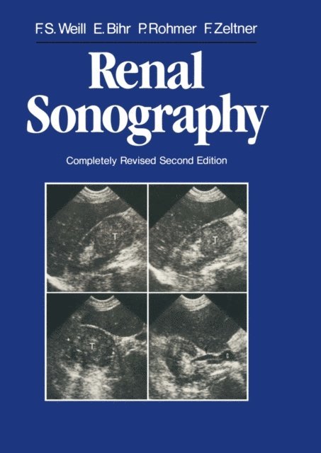
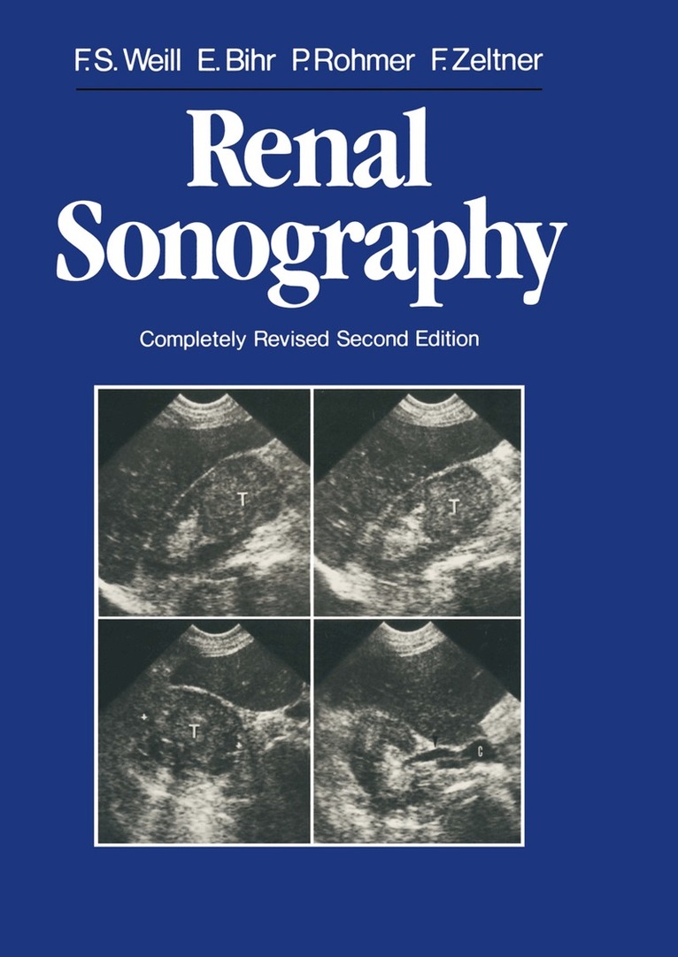
Renal sonography forms a basic part of routine diagnostic strategy. This textbook summarizes eighteen years of experience in diagnostic ultrasound. We want it to serve as a guide to both imagers and mere consumers of information. That is why we shall emphasize practical advice and diagnostic pitfalls; it is also why we shall often deal with the relations of sonography with other diagnostic procedures, which it may complement or replace, precede or follow, the purpose being to achieve efficiency at low cost. We shall limit our subject matter to the kidney itself and the neighboring retroperitoneal compartments, dealing only briefly with the lower urinary tract, which requires specialized procedures. We devoted considerable space to renal sonography in our book Clinical Atlas of Ultrasonic Radiography, published in 1973. Since then, nothing has changed and everything has changed. Nothing, because even then the differential diagnosis between a solid and a cystic mass, the etiologic diagnosis of a nonsecreting kidney, and the positive diagnosis of a traumatic juxtarenal hematoma were quite reliable, making possible drastic reductions in the indications for instrumental roentgenologic examinations. Everything, be cause improved resolution and grey scale imaging (already partially achieved in 1973, thanks to real time) have profoundly refined both anatomic and pathologic ultrasonic studies. And now high resolution real time imaging has revolutionized renal examination techniques, whereas Doppler is entering routine ultrasonic diagnosis.
Häftad, Engelska, 2013
1 349 kr
Skickas inom 10-15 vardagar
Renal sonography forms a basic part of routine diagnostic strategy. This textbook summarizes eighteen years of experience in diagnostic ultrasound. We want it to serve as a guide to both imagers and mere consumers of information. That is why we shall emphasize practical advice and diagnostic pitfalls; it is also why we shall often deal with the relations of sonography with other diagnostic procedures, which it may complement or replace, precede or follow, the purpose being to achieve efficiency at low cost. We shall limit our subject matter to the kidney itself and the neighboring retroperitoneal compartments, dealing only briefly with the lower urinary tract, which requires specialized procedures. We devoted considerable space to renal sonography in our book Clinical Atlas of Ultrasonic Radiography, published in 1973. Since then, nothing has changed and everything has changed. Nothing, because even then the differential diagnosis between a solid and a cystic mass, the etiologic diagnosis of a nonsecreting kidney, and the positive diagnosis of a traumatic juxtarenal hematoma were quite reliable, making possible drastic reductions in the indications for instrumental roentgenologic examinations. Everything, be cause improved resolution and grey scale imaging (already partially achieved in 1973, thanks to real time) have profoundly refined both anatomic and pathologic ultrasonic studies. And now high resolution real time imaging has revolutionized renal examination techniques, whereas Doppler is entering routine ultrasonic diagnosis.
E-bok
PDF, Tyska, 2013603 kr
Läs direkt efter köp
Dieses Buch bietet, kurzgefaßt und übersichtlich in 13 Kapital gegliedert, eine umfassende Einführung in die Anwendung der Computertomographie im Thorakal- und Abdominalbereich; die im Text gegebenen Anleitungen werden großzügig durch didaktisch hervorragendes Bildmaterial illustriert. Das Buch wendet sich nicht nur an Ärzte, die in der radiologischen Fortbildung stehen, sondern ist darüber hinaus auch für zuweisende Ärzte anderer Fachrichtungen (Allgemeinärzte, Internisten, Chirurgen) von ganz besonderem Interesse, da es knapp und kompetent über die Indikationen und diagnostischen Möglichkeiten der Computertomographie informiert.
E-bok
PDF, Engelska, 20121 459 kr
Läs direkt efter köp
During the 1970s ultrasonography developed along different paths in continental Eu rope and the English-speaking world. Whereas static imaging dominated the United Kingdom and North America, real-time examination performed by physicians was the norm in the Federal Republic of Germany and France. Francis Weill was uniquely able to bridge the gap between the two schools since he employed both techniques extensively. He made many important contributions to ab dominal ultrasonography during that decade, including the first descriptions of peri pancreatic vascular anatomy, the sonographic diagnostic features of obstructive jaun dice, and the localization of intraperitoneal fluid collections. During the past 20 years he has been working on an evolving textbook of gastroin testinal ultrasonography. This edition in English is the latest fruit of that effort. There are many textbooks of ultrasonography but very few are the work of a single author with a systematic, consistent, and thoroughly organized approach. Moreover, a work that has been crafted and refined by 20 years of clinical experience is certainly unique in this field. My relationship to this book began with the first English edition which I helped to translate from French. The current volume is a revised version of the most recent French edition, a book that we have found to be an invaluable reference in our de partment of radiological ultrasound for anyone who can manage a bit of French. I .