Herwig Imhof – författare
895 kr
Skickas inom 5-8 vardagar
2 516 kr
Skickas inom 5-8 vardagar
1 905 kr
Läs direkt efter köp
Lavishly illustrated with hundreds of superb MR images and CT scans
The diagnosis of trauma to the spine -- where the slightest oversight may have catastrophic results -- requires a thorough grasp of the spectrum of resultant pathology as well as the imaging modalities used in making an accurate diagnosis. In Spinal Trauma, the internationally renowned team of experts provides a comprehensive, cutting-edge exposition of the current vital role of imaging in the diagnosis and treatment of injuries to the axial skeleton. Beginning with a valuable clinical perspective of spinal trauma, the book offers the reader a unique overview of the biomechanics underlying the beautifully illustrated pathology of cervical trauma.
Acute trauma topics include:
Optimization of imaging modalities Malalignment -- signs and significance Vertebral fractures -- detection and implications Classification of thoraco-lumbar fractures -- rationale and relevance Neurovascular injuryDistilling decades of clinical and teaching expertise, the contributors further discuss the current role of imaging in special focus topics, which include:
The pediatric spine Sports injuries The rigid spine Trauma in the elderly Vertebral collapse, benign and malignant Spinal trauma therapy Vertebral fractures and osteoporosis Neuropathic spineAll throughout the book, the focus is on understanding the injury, and its implications and complications, through "an imaging approach." Complete with hundreds of superb MR images and CT scans, and clear full-color drawings, the authors conclude with a look into the future, defining clinical trends and research directions. Spinal Trauma -- with its broad scope, practical imaging approach, and current focus -- is designed to enhance confidence and accuracy, making it essential reading for clinicians and radiologists at all levels.
952 kr
Läs direkt efter köp
Due to the multitude of bone and joint disorders and their symptomatic similarities, establishing a differential diagnosis is often problematic in daily practice. This book offers invaluable help by showing the diagnostic effectiveness of multimodality imaging across the entire spectrum of bone and joint disorders.
Each clinical entity is presented as a unit, with succinct text on the left and high-quality, labeled images on the right. A consistent structure featuring pathology, clinical findings, radiology, nuclear medicine, MRI, and differential diagnosis offers quick access to the information you need for any given bone, joint, or soft tissue disease.
More than 1,300 high-quality radiologic images and two-color drawings that allow you to visualize each disorder. Key information presented in just 404 pages, saving you the time and inconvenience of wading through large texts. Useful tables summarizing radiologic findings for each disorder.
All-inclusive coverage, with in-depth treatment of such important areas as trauma.
710 kr
Läs direkt efter köp
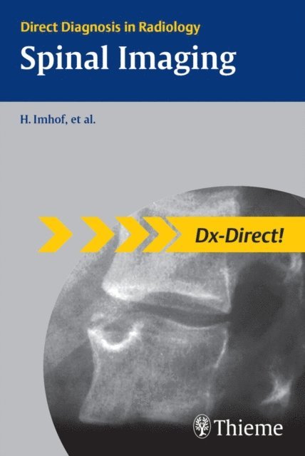
Dx-Direct is a series of eleven Thieme books covering the main subspecialties in radiology. It includes all the cases you are most likely to see in your typical working day as a radiologist. For each condition or disease you will find the information you need -- with just the right level of detail.
Whether you are a resident or a trainee, preparing for board examinations or just looking for a superbly organized reference: Dx-Direct is the high-yield choice for you!
The series covers the full spectrum of radiology subspecialties including:
Brain Gastrointestinal Cardiac Breast Urogenital Vascular Spinal Head and Neck Musculoskeletal Pediatric ThoracicDx-Direct gets to the point: Definitions, Epidemiology, Etiology, and Imaging Signs Typical Presentation, Treatment Options, Course and Prognosis Differential Diagnosis, Tips and Pitfalls, and key References ... all combined with high-quality diagnostic images.
3 708 kr
Läs direkt efter köp
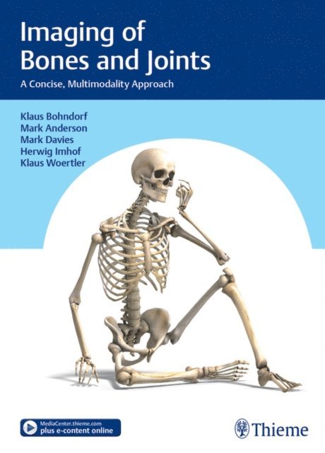
Imaging of Bones and Joints: A Concise, Multimodality Approach
This book is unique. It will guide you through the essentials of musculoskeletal imaging using a multimodality approach. Organized by categories of musculoskeletal disorders, it uses a "findings within-the-image" method to help you identify the typical imaging features of each condition.
As a comprehensive reference compiled by well-known specialists in the field, it is useful for both practicing radiologists and those in training.
Focus on the essentials
Provides a solid foundation of what the radiologist needs to know when interpreting musculoskeletal imaging studies, including the indications for when to use various imaging modalities.
"Findings within the image"
An excellent presentation method for learning to interpret bone and joint images.
Find it quickly
In addition to a detailed text and high-quality images, important points are summarized in boxes, tables, and illustrative figures for quick reference.
Extra features are included on the Thieme MediaCenter
An additional 338 images along with supplemental text and references are provided online on the Thieme MediaCenter.
Special Features
All chapters are written by leading international authors.A comprehensive, multimodality approach is used.Over 2100 brilliant, state-of-the-art images are provided, including a multitude of MR images.