Myrtle A. Davis – författare
Visar alla böcker från författaren Myrtle A. Davis. Handla med fri frakt och snabb leverans.
3 produkter
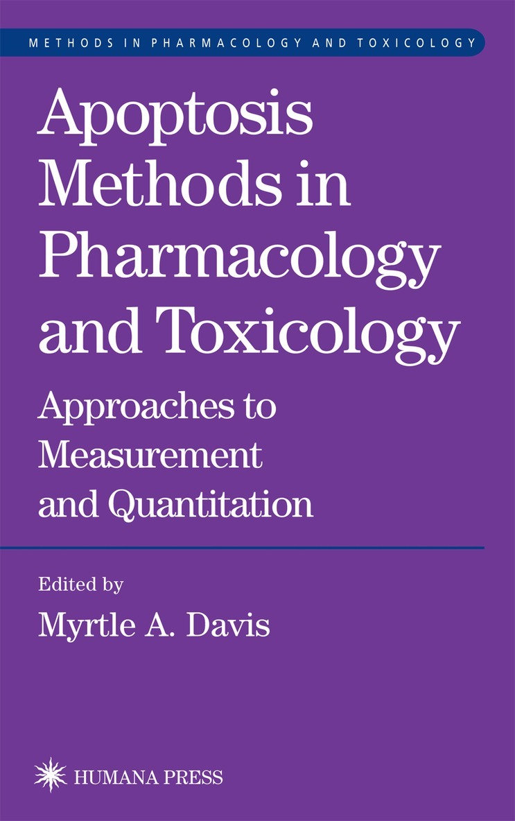
Apoptosis Methods in Pharmacology and Toxicology
Approaches to Measurement and Quantification
Inbunden, 2002
1131 kr
Lägg i varukorg
Dr. Myrtle A. Davis has assembled a panel of cutting-edge scientists to describe their best methods for detecting, illuminating, and quantifying apoptotic mechanisms in a way that is useful for the design of toxicology and pharmacology studies. These state-of-the-art techniques include flow …
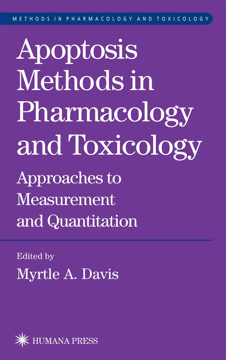
Apoptosis Methods in Pharmacology and Toxicology
Approaches to Measurement and Quantification
E-bok, 2007
1502 kr
Lägg i varukorg
Dr. Myrtle A. Davis has assembled a panel of cutting-edge scientists to describe their best methods for detecting, illuminating, and quantifying apoptotic mechanisms in a way that is useful for the design of toxicology and pharmacology studies. These state-of-the-art techniques include flow …
Apoptosis Methods in Pharmacology and Toxicology
Approaches to Measurement and Quantification
Häftad, 2011
1131 kr
Lägg i varukorg
Dr. Myrtle A. Davis has assembled a panel of cutting-edge scientists to describe their best methods for detecting, illuminating, and quantifying apoptotic mechanisms in a way that is useful for the design of toxicology and pharmacology studies. These state-of-the-art techniques include flow …