Pierre Grangeat – författare
Visar alla böcker från författaren Pierre Grangeat. Handla med fri frakt och snabb leverans.
6 produkter
6 produkter
E-bok
PDF, Engelska, 20102 830 kr
Läs direkt efter köp
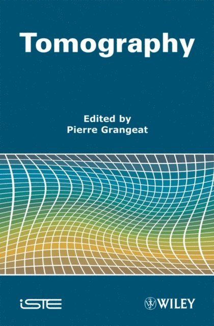
The principle of tomography is to explore the structure and composition of objects non-destructively along spatial and temporal dimensions, using penetrating radiation, such as X- and gamma-rays, or waves, such as electromagnetic and acoustic waves. Based on computer-assisted image reconstruction, tomography provides maps of parameters that characterize the emission of the employed radiation or waves, or their interaction with the examined objects, for one or several cross-sections. Thus, it gives access to the inner structure of inert objects and living organisms in their full complexity. In this book, multidisciplinary specialists explain the foundations and principles of tomographic imaging and describe a broad range of applications. The content is organized in five parts, which are dedicated to image reconstruction, microtomography, industrial tomography, morphological medical tomography and functional medical tomography.
Inbunden, Engelska, 1996
1 119 kr
Skickas inom 10-15 vardagar
This book contains twenty-one selected papers based on communications presented at the "Third International Meeting on Fully Three-Dimensional Image Reconstruction in Radiology and Nuclear Medicine", held July 4-6, 1995 at Domaine d'Aix- Marlioz, Aix-les-Bains, France. 3D tomographic imaging systems based on ionising radiations tend to use 2D detectors in order to improve the radiation detection efficiency. Then, fully 3D image reconstruction algorithms are required to recover the 3D image of the region of interest. These systems include 3D radiology, 3D X-ray computerised tomography, single photon emission computerised tomography (SPECT), and positron emission tomography (PET). The material is this text divided into four parts covering the following topics: cone-beam and new geometries reconstruction, SPECT quantitation, patient motion and gated SPECT, and PET quantitation and reconstruction.Aud ience:This work will be of interest to scientists, physicists and physicians seeking new information and insight in the on-going research work in this expanding field.
E-bok
Engelska, 20132 932 kr
Läs direkt efter köp
The principle of tomography is to explore the structure and composition of objects non-destructively along spatial and temporal dimensions, using penetrating radiation, such as X- and gamma-rays, or waves, such as electromagnetic and acoustic waves. Based on computer-assisted image reconstruction, tomography provides maps of parameters that characterize the emission of the employed radiation or waves, or their interaction with the examined objects, for one or several cross-sections. Thus, it gives access to the inner structure of inert objects and living organisms in their full complexity. In this book, multidisciplinary specialists explain the foundations and principles of tomographic imaging and describe a broad range of applications. The content is organized in five parts, which are dedicated to image reconstruction, microtomography, industrial tomography, morphological medical tomography and functional medical tomography.
Inbunden, Engelska, 2009
2 573 kr
Skickas inom 11-20 vardagar
The principle of tomography is to explore the structure and composition of objects non-destructively along spatial and temporal dimensions, using penetrating radiation, such as X- and gamma-rays, or waves, such as electromagnetic and acoustic waves. Based on computer-assisted image reconstruction, tomography provides maps of parameters that characterize the emission of the employed radiation or waves, or their interaction with the examined objects, for one or several cross-sections. Thus, it gives access to the inner structure of inert objects and living organisms in their full complexity. In this book, multidisciplinary specialists explain the foundations and principles of tomographic imaging and describe a broad range of applications. The content is organized in five parts, which are dedicated to image reconstruction, microtomography, industrial tomography, morphological medical tomography and functional medical tomography.
Del 4 - Computational Imaging and Vision
Three-Dimensional Image Reconstruction in Radiology and Nuclear Medicine
Häftad, Engelska, 2010
1 123 kr
Skickas inom 10-15 vardagar
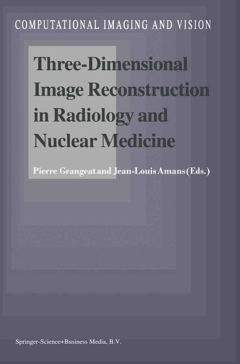
This book contains a selection of communications presented at the Third International Meeting on Fully Three-Dimensional Image Reconstruction in Radiology and Nuclear Medicine, held 4-6 July 1995 at Domaine d' Aix-Marlioz, Aix-Ies-Bains, France. This nice resort provided an inspiring environment to hold discussions and presentations on new and developing issues. Roentgen discovered X-ray radiation in 1895 and Becquerel found natural radioactivity in 1896 : a hundred years later, this conference was focused on the applications of such radiations to explore the human body. If the physics is now fully understood, 3D imaging techniques based on ionising radiations are still progressing. These techniques include 3D Radiology, 3D X-ray Computed Tomography (3D-CT), Single Photon Emission Computed Tomography (SPECT), Positron Emission Tomography (PET). Radiology is dedicated to morphological imaging, using transmitted radiations from an external X-ray source, and nuclear medicine to functional imaging, using radiations emitted from an internal radioactive tracer. In both cases, new 3D tomographic systems will tend to use 2D detectors in order to improve the radiation detection efficiency. Taking a set of 2D acquisitions around the patient, 3D acquisitions are obtained. Then, fully 3D image reconstruction algorithms are required to recover the 3D image of the body from these projection measurements.
E-bok
PDF, Engelska, 20131 459 kr
Läs direkt efter köp
This book contains a selection of communications presented at the Third International Meeting on Fully Three-Dimensional Image Reconstruction in Radiology and Nuclear Medicine, held 4-6 July 1995 at Domaine d'' Aix-Marlioz, Aix-Ies-Bains, France. This nice resort provided an inspiring environment to hold discussions and presentations on new and developing issues. Roentgen discovered X-ray radiation in 1895 and Becquerel found natural radioactivity in 1896 : a hundred years later, this conference was focused on the applications of such radiations to explore the human body. If the physics is now fully understood, 3D imaging techniques based on ionising radiations are still progressing. These techniques include 3D Radiology, 3D X-ray Computed Tomography (3D-CT), Single Photon Emission Computed Tomography (SPECT), Positron Emission Tomography (PET). Radiology is dedicated to morphological imaging, using transmitted radiations from an external X-ray source, and nuclear medicine to functional imaging, using radiations emitted from an internal radioactive tracer. In both cases, new 3D tomographic systems will tend to use 2D detectors in order to improve the radiation detection efficiency. Taking a set of 2D acquisitions around the patient, 3D acquisitions are obtained. Then, fully 3D image reconstruction algorithms are required to recover the 3D image of the body from these projection measurements.