2 333 kr
Skickas inom 5-8 vardagar
2 333 kr
Skickas inom 5-8 vardagar
3 108 kr
Läs direkt efter köp
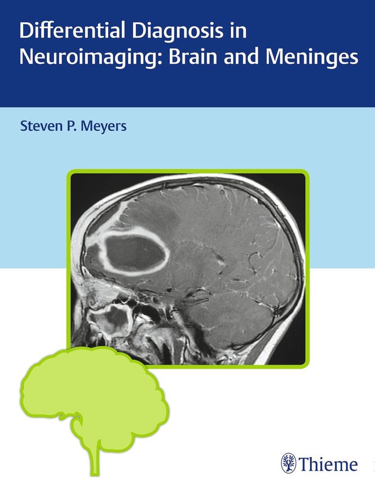
Authored by renowned neuro-radiologist Steven P. Meyers, Differential Diagnosis in Neuroimaging: Brain and Meninges is a stellar guide for identifying and diagnosing brain pathologies based on location and neuroimaging results. The succinct text reflects more than 25 years of hands-on experience gleaned from advanced training and educating residents and fellows in radiology, neurosurgery, and neurology. The high-quality MRI, CT, PET, PET/CT, conventional angiography, and X-ray images have been collected over Dr. Meyers''s lengthy career, presenting an unsurpassed visual learning tool.
The distinctive ''three-column table plus images'' format is easy to incorporate into clinical practice, setting this book apart from larger, disease-oriented radiologic tomes. The layout enables readers to quickly recognize and compare abnormalities based on high-resolution images.
Key Highlights
Tabular columns organized by anatomical abnormality include brain imaging findings and a summary of key clinical data that correlates to the imagesComprehensive imaging of the brain, ventricles, meninges, and neurovascular system in both children and adults, including congenital/developmental anomalies and acquired diseaseMore than 1,900 figures illustrate the radiological appearance of intracranial lesions, masses, neurodegenerative disorders, ischemia and infarction, and moreThis visually rich resource is a must-have diagnostic tool for radiologists, neurosurgeons, and neurologists, and residents and fellows. The highly practical format makes it ideal for daily rounds, as well as a robust study guide for physicians preparing for board exams.
3 108 kr
Läs direkt efter köp
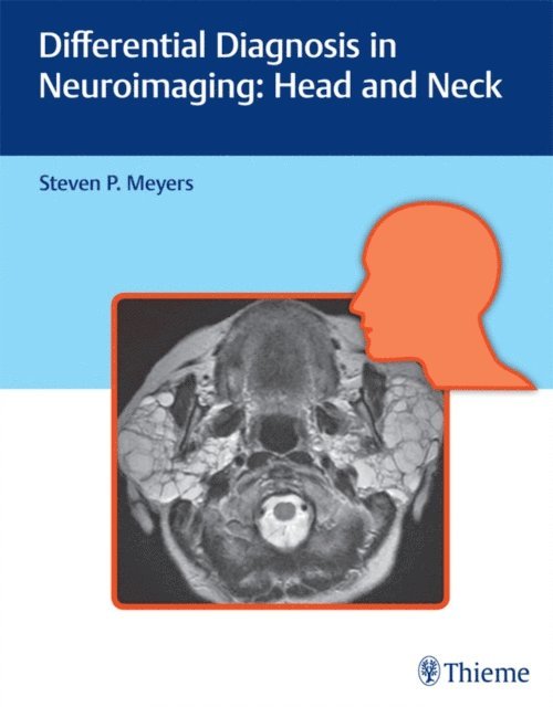
Authored by renowned neuro-radiologist Steven P. Meyers, Differential Diagnosis in Neuroimaging: Head and Neck is a stellar guide for identifying and diagnosing head and neck disease based on location and neuroimaging results. The succinct text reflects more than 25 years of hands-on experience gleaned from advanced training and educating residents and fellows in radiology, neurosurgery, and otolaryngology. The high-quality MRI and CT scans have been collected over Dr. Meyers''s lengthy career, presenting an unsurpassed visual learning tool.
The distinctive ''three-column table plus images'' format is easy to incorporate into clinical practice, setting this book apart from larger, disease-oriented radiologic tomes. This layout enables readers to quickly recognize and compare abnormalities based on more than 1,500 high-resolution images. Chapters cover skull imaging, temporal bone imaging, orbital imaging, paranasal imaging, suprahyoid neck imaging, and infrahyoid neck imaging, for a full spectrum of head and neck pathologies.
Key Highlights
Tabular columns organized by anatomical abnormality include neck, facial, and skull based imaging findings and a summary of key clinical data that correlate to the imagesCongenital/developmental and acquired abnormalities including solitary or multiple orbital lesions; and solitary, multifocal, or diffuse sinonasal diseaseAbnormalities of the skull, craniovertebral junction, tempormandibular joint, infrahyoid neck, anterior and posterior cervical space, perivertebral space, and brachial plexusThis visually rich resource is a must-have diagnostic tool for residents, fellows, and practitioners in radiology, otolaryngology-head and neck surgery, and neurosurgery. The highly practical format makes it ideal for daily rounds, as well as a robust study guide for physicians preparing for board exams.
2 590 kr
Läs direkt efter köp
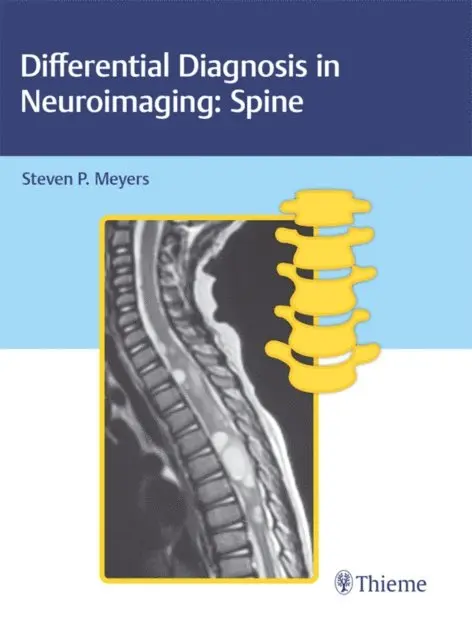
A unique, easy-to-use, and definitive guide to diagnostic spine imaging
Authored by renowned neuro-radiologist Steven P. Meyers, Differential Diagnosis in Neuroimaging: Spine is a stellar guide for identifying and diagnosing cervical, thoracic, lumbar, and sacral spine anomalies based on location and neuroimaging results. The succinct text reflects more than 25 years of hands-on experience gleaned from advanced training and educating residents and fellows in radiology, neurosurgery, and orthopaedic surgery. The high-quality MRI, CT, and X-ray images have been collected over Dr. Meyers''s lengthy career, presenting an unsurpassed visual learning tool.
The distinctive ''three-column table plus images'' format is easy to incorporate into clinical practice, setting this book apart from larger, disease-oriented radiologic tomes. This layout enables readers to quickly recognize and compare abnormalities based on high-resolution images.
Key Highlights
Tabular columns organized by anatomical abnormality include imaging findings and a summary of key clinical data that correlates to the imagesCongenital/developmental abnormalities, spinal deformities, and acquired pathologies in both children and adultsLesions organized by region including dural, intradural extramedullary, extra-dural, and sacrumMore than 600 figures illustrate the radiological appearance of spinal tumors, lesions, deformities, and injuriesSpinal cord imaging for the diagnosis of intradural intramedullary lesions and spinal traumaThis visually rich resource is a must-have diagnostic tool for trainee and practicing radiologists, neurosurgeons, neurologists, physiatrists, and orthopaedic surgeons who specialize in treating spine-related conditions. The highly practical format makes it ideal for daily rounds, as well as a robust study guide for physicians preparing for board exams.
3 463 kr
Läs direkt efter köp
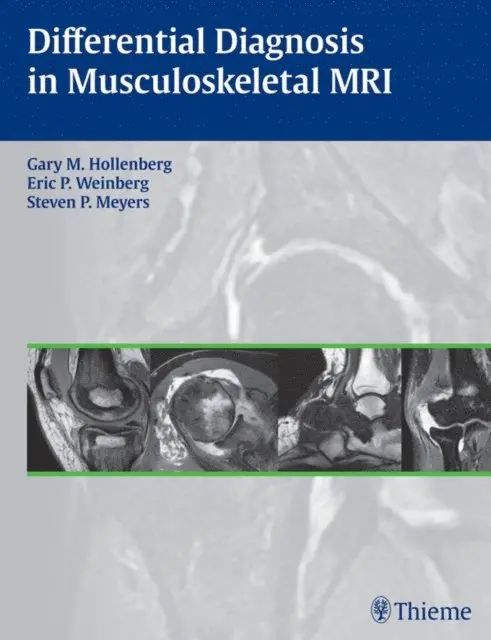
The definitive guide to MRI for musculoskeletal abnormalities
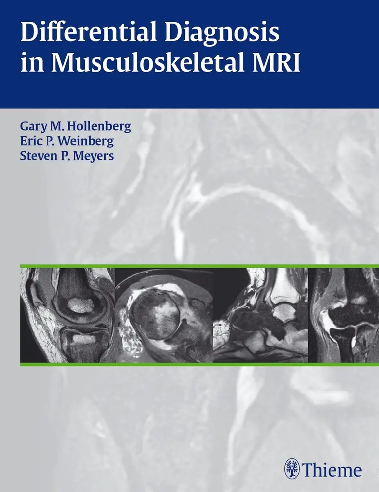
Differential Diagnosis in Musculoskeletal MRI is a unique desk reference offering extensive descriptions of MRI findings that enable radiologists to more easily diagnose a wide range of musculoskeletal conditions. The authors have placed the most essential content covering etiologic, clinical, and pathological aspects in tabular format to assist readers in quickly and efficiently obtaining the information they need.
Features:
More than 2,000 state-of-the-art images covering the features for both common and rare musculoskeletal conditionsAuthors provide a practical approach to MRI evaluation and differential diagnosis selectionDifferential diagnosis tables are based on anatomic locations of lesionsThis practical reference is an invaluable tool that practicing radiologists, residents, and fellows will use to diagnose both common and rare lesions as well as other disorders of the musculoskeletal system. It is an excellent guide for use in daily practice as well as a comprehensive exam preparation tool.