• Fri frakt över 249 kr
  • •
  • Snabba leveranser
  • •
  • Billiga böcker
Kundservice

Du är på sajten för privatpersoner.

Företag, bibliotek eller offentlig verksamhet?

Du handlar på classic.bokus.com, där alla dina funktioner finns intakta.
Till classic.bokus.com
Bokus logotyp. Gå till startsidan.
  • Erbjudanden
  • Nyheter
  • Student
  • Topplistor
  • Barn & ungdom
  • Bokus Play
  • E-böcker
  • Pocketböcker
  • Spel & pussel

10% rabatt på allt med kod: NYSTART10 →

Sidfot

Mina sidor

    Hjälp

    • Kundservice
    • Vanliga frågor och svar
    • Frakt och leverans
    • Retur vid ångerrätt
    • Reklamera vara
    • Betalning
    • Köpvillkor
    • Allmänna villkor
    • Information om webbplatsens tillgänglighet

    Om Bokus

    • Om oss
    • Pressrum
    • För studenter
    • För företag
    • För bibliotek och offentlig verksamhet
    • För leverantörer
    • Hållbarhet

    Populärt

    • Aktuella erbjudanden
    • Presentkort
    • Studentlitteratur
    • Nya böcker
    • Topplistor
    • Signerade böcker
    • Engelska böcker

    Inspiration

    • Boktips
    • BookTok
    • Populära bokserier
    • Barnbokskaraktärer
    • Populära författare
    Logotyp för Bokus
    Följ oss på Facebook (extern länk)Följ oss på Instagram (extern länk)Följ oss på YouTube (extern länk)Följ oss på TikTok (extern länk)
    bokus @ CookiesAnpassa cookiesIntegritetspolicyKöpvillkor
    Till Citymail hemsida (extern länk)Till Budbee hemsida (extern länk)Till Postnord hemsida (extern länk)Till Schenker hemsida (extern länk)Till Early Bird hemsida (extern länk)Till Walleys hemsida (extern länk)
    1. Medicin
    2. Omvårdnad och medicinska stödfunktioner
    3. Rehabilitering

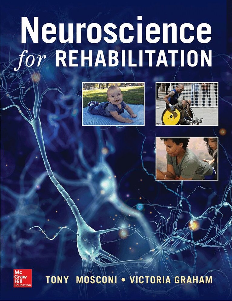
    Neuroscience for Rehabilitation

    AvTony Mosconi,Victoria Graham

    Häftad, Engelska, 2018

    1 116 kr

    Beställningsvara. Skickas inom 3-6 vardagar. Fri frakt över 249 kr.

    Beskrivning

    Publisher's Note: Products purchased from Third Party sellers are not guaranteed by the publisher for quality, authenticity, or access to any online entitlements included with the product.


    The first neuroanatomy text written specifically for physical therapy students

    Written by recognized experts in human nervous system development, Neuroanatomy for Rehabilitation provides physical therapy students with a thorough understanding of the anatomical localization of brain function. Approximately 200 line illustrations and photographs teach students how to accurately interpret the wealth of new human brain images now available.

    The text opens with an informative section that discusses the structural and functional organization of the nervous system and includes coverage of functional neuroanatomy and response to injury. This is followed by sections covering:

    • Vascular Supply of the Central Nervous System (arterial supply, venous drainage, response to injury)
    • Cellular Organization of the Nervous System
    • Development of the Central Nervous System
    • Functional Neuroanatomy by Ascending Region
    • The Brainstem, Cranial Nerves and Visual Pathways
    • The Cerebellum and Basal Ganglia
    • Diencephalon
    • The Cortex

    Each section opens with a case study and an overview of key concepts, and concludes with case discussion and review questions. No other text provides physical therapy students and instructors with such relevant, authoritative, and well-written material designed to enhance understanding of the human nervous system as Neuroanatomy for Rehabilitation.

    Produktinformation

    • Utgivningsdatum:2018-03-04
    • Mått:221 x 279 x 15 mm
    • Vikt:771 g
    • Format:Häftad
    • Språk:Engelska
    • Antal sidor:336
    • Förlag:McGraw-Hill Education
    • ISBN:9780071828888

    Utforska kategorier

    • Rehabilitering inom Medicin
    • Fysioterapi inom Medicin

    Innehållsförteckning

    • Neuroanatomy for Physical TherapistsDraft Table of ContentsI. Introduction to the Nervous SystemA. Neuronal CytologyB. Neuronal Signaling (excitability, membrane, action potential, AP propagation and fiber diameter, synapses)C. Functional Organization of the NS1. PNS (nerve roots, spinal Nn, plexuses, peripheral nerves, sensory endings, motor endplate, Wallerian degeneration, sprouting/regeneration, demyelinating diseases)2. CNS a. Support cellsb. Pathways, tracts, fasciculi, funiculi, columns, lemniscic. Spinal cord (topology, internal anatomy)d. Brainstem (topology, internal anatomy)e. Diencephalon (topology, internal anatomy)f. Forebrain (topology, internal anatomy)g. Cerebral cortex topography and functional specialization3. ANS (sympathetics, parasympathetics, pre- and postganglionics, autonomic ganglia)II. Blood and CSF CirculationA. Carotid and Vertebral Aa (circle of Willis)B. Spinal Cord and Brainstem Blood SupplyC. Forebrain Blood SupplyD. Meninges And Brain CoveringsE. CSF Production And Circulation (hydrocephalus)III. Development of the CNSA. Embryonic Brain (dysraphic and myeloschistic defects)B. Juvenile Brain (CP, MS)C. Mature Brain (ALS)D. Aged Brain (Alzheimer’s, dementia, Parkinson’s)E. Critical Periods and NeuroplasticityIV. Functional Neuroanatomy by RegionA. Spinal Cord1. PNS-CNS junction2. Long ascending somatosensory pathwaysa. Dorsal column-medial lemniscusb. Anterolateral system (Spinothalamic tract)3. Long descending somatic motor pathwaysa. Corticospinal tractb. UMN-LMNc. Reflexesd. Spasticity4. Brown-Séquard syndromeB. Brainstem1. Long pathwaysa. DC-MLb. ALSc. CSTd. Corticobulbar (Corticonuclear) Tract (unilateral lesion, facial paresis)2. Medullaa. CNn nuclei (XII, X, IX, VIII, VII) and peripheral nervesb. Lateral medullary syndromec. Medial medullary syndrome – inferior alternating hemiplegia3. Ponsa. CNn nuclei (VI, V) and peripheral nervesb. Locked-in syndromec. Middle alternating hemiplegiad. Acoustic neuroma4. Midbraina. CNn nuclei (IV, III) and peripheral nervesb. Weber’s syndrome – superior alternating hemiplegia 5. Diencephalona. Thalamusb. Internal capsulec. Thalamic and capsular lesionsC. Cerebellum1. Topology, internal anatomy2. Cerebellar cortex circuitry3. Afferent input4. Efferent output5. Motor learning6. Balance7. Ataxia, intention tremor, ipsilateral deficitsD. Forebrain1. Basal Ganglia (Basal Nuclei)a. Component nucleib. Circuitryc. Parkinson’s diseased. Huntington’s choreae. Contralateral hemiballismus2. Cerebruma. Cerebral cortex architectonics (6 layers, input and output layers, regional functional differentiation)b. Fiber systems (superior longitudinal fasciculus, arcuate fasciculus, corpus callosum, anterior commissure, corona radiata, optic radiations)c. Gyri and sulci1) Topology2) Functional differentiationd. Deficits – TBI, CVAV. Vestibular SystemA. Peripheral AnatomyB. Brainstem AnatomyC. Circuitry (local, ascending, and descending pathways, MLF)D. Vestibulo-ocular ReflexesE. Lesions (vertigo, nystagmus)F. Therapeutic ManipulationVI. Visual SystemA. Anatomy (eye through visual cortex) and connectivityB. Sensory Function1. Construction of the visual field2. Deficits (blindness, hemianopsia, quadrantopia, complete blindness, pituitary tumor, macular degeneration/sparing)C. Motor Control of the Visual System1. Eye movements (CN III palsy, CN IV palsy, CN VI lateral rectus and lateral gaze palsies, internuclear ophthalmoplegia)2. Eye movement reflexes (VOR, oculocephalic reflex, doll’s eye sign)3. Pupillary reflexes D. Vestibulo-optic Interactions – functional and therapeutic implicationsVII. Reticular FormationA. Anatomy and ConnectivityB. NeurotransmittersC. Reticular Activating System1. Consciousness, arousal, sleep-wake2. Vital functions3. Descending pain modulation4. Lesions (infratentorial herniation, decerebrate/decorticate posturing)5. Therapeutic manipulationVIII. Limbic SystemA. Anatomy and Connectivity (hippocampal formation, Papez circuit)B. Learning, Memory, EmotionsC. Olfaction and the Limbic SystemC. Lesions (temporal lobe epilepsy, anterograde amnesia, Alzheimer’s, dementia, Korsakoff syndrome, Kluver-Bucy)IX. Hypothalamus and the ANSA. Anatomy and Connectivity of the HypothalamusB. Central Autonomic Structures1. Sympathetic (spinal cord lateral horn, preganglionic neurons)2. Parasympathetic (cranial and sacral preganglionic neurons)C. Peripheral Autonomics1. Sympathetic (ganglia, postganglionics)2. Parasympathetic (ganglia, postganglionics)D. Lesions (hypothalamic syndrome, complex regional pain syndrome) X. PainA. Peripheral nociceptors – Primary and secondary hyperalgesiaB. Spinal cord dorsal horn (substantia gelatinosa) and spinothalamic tractC. Reticular formation, PAG, and the hypothalamus – Descending pain modulationD. Chronic PainE. Maladaptive cortical remappingAppendix 1Neuroimaging (assessing images and diagnosing lesion)Appendix 2The complete neurological exam Atlas