Atsushi Iriki – författare
Visar alla böcker från författaren Atsushi Iriki. Handla med fri frakt och snabb leverans.
5 produkter
5 produkter
Inbunden, Engelska, 2019
2 239 kr
Skickas inom 10-15 vardagar
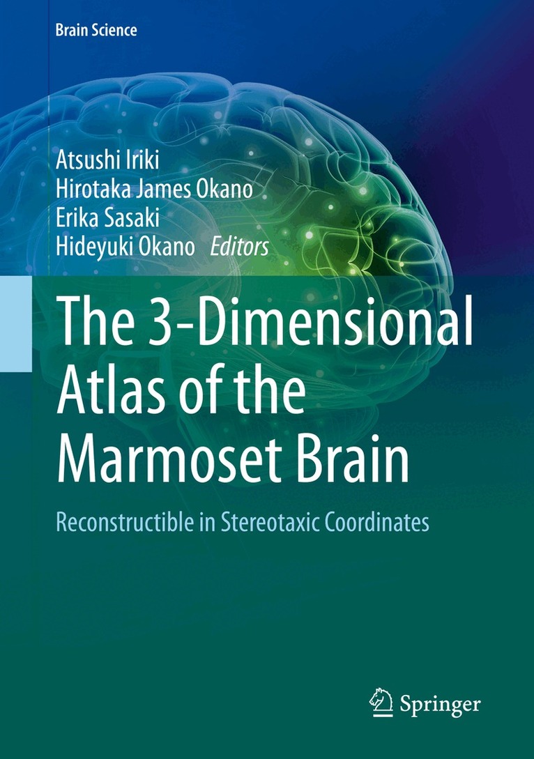
This book provides accurate, comprehensive, and convenient reference for usages of the “freely rotatable three dimensional combined Nissl-stained and MRI digital data of the marmoset brain”. The key features of the original 3D digital data and of this atlas are: 1. The original digital datasets are freely rotatable in three dimensions, thus expected to be useful for any disciplines and anatomical interest, using any coordinate system, 2. Combined Nissl stained and MRI images are obtained from the same marmoset, to allow cross-modality matched references for multiple usages, 3. 86 Horizontal Series of Images with Neurosurgical Plane (based on the actual data), with more accuracy and resolution (Chapter 2) than the web-based digital images, 4. 32 Coronal Series of Images with Neurosurgical Plane (reproduced from the brain model) (Chapter 3), 5. 10 Parasagittal Series of Images with Neurosurgical Plane (reproduced from the brain model) (Chapter 4), 6. 3 Omnidirectionally Sliceable Planes (reproduced from the brain model) (Chapter 5), 7. In order to provide higher resolution structures to match systematic accuracy for supplementation of the digital data on the website, additional information are included. They are: 1) Nomenclature, 2) List of Brain Structures in Hierarchical Order, 3) Index of Abbreviations, together with 143 useful Bibliographic References list as of 2016, 8. Horsley-Clarke’s stereotaxic coordinates were adopted in the present atlas.
E-bok
Engelska, 20182 848 kr
Läs direkt efter köp
This book provides accurate, comprehensive, and convenient reference for usages of the “freely rotatable three dimensional combined Nissl-stained and MRI digital data of the marmoset brain”. The key features of the original 3D digital data and of this atlas are: 1. The original digital datasets are freely rotatable in three dimensions, thus expected to be useful for any disciplines and anatomical interest, using any coordinate system, 2. Combined Nissl stained and MRI images are obtained from the same marmoset, to allow cross-modality matched references for multiple usages, 3. 86 Horizontal Series of Images with Neurosurgical Plane (based on the actual data), with more accuracy and resolution (Chapter 2) than the web-based digital images, 4. 32 Coronal Series of Images with Neurosurgical Plane (reproduced from the brain model) (Chapter 3), 5. 10 Parasagittal Series of Images with Neurosurgical Plane (reproduced from the brain model) (Chapter 4), 6. 3 Omnidirectionally Sliceable Planes (reproduced from the brain model) (Chapter 5), 7. In order to provide higher resolution structures to match systematic accuracy for supplementation of the digital data on the website, additional information are included. They are: 1) Nomenclature, 2) List of Brain Structures in Hierarchical Order, 3) Index of Abbreviations, together with 143 useful Bibliographic References list as of 2016, 8. Horsley-Clarke’s stereotaxic coordinates were adopted in the present atlas.
Inbunden, Engelska, 2018
1 123 kr
Skickas inom 10-15 vardagar
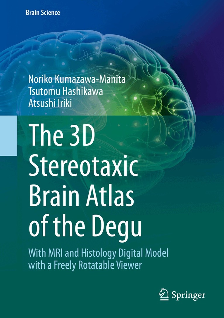
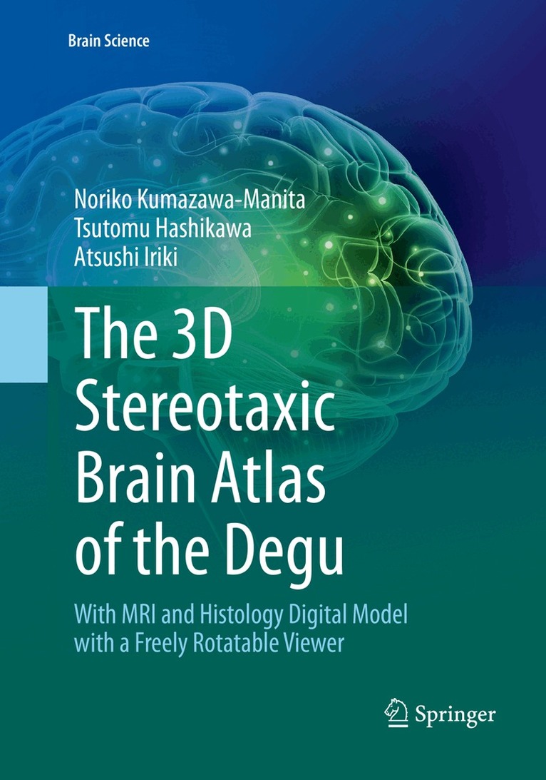
This book is the first digital atlas of the degu brain with microscopic features simultaneously in Nissl sections and magnetic resonance imaging (MRI). As an experimental animal model, the degu contributes to a variety of medical research fields in diabetes, hyperglycemia, pancreatic function, and adaptation to high altitude, among others. Recently the degu has gained increasing importance in the field of neuroscience, particularly in studies evaluating the relationship between sociality and cognitive brain functions, and in studies pertaining to the evolutional aspects of the acquisition of tool-use abilities. Furthermore, aging-related brain dysfunction in humans can be studied using this animal model in addition to mammals with much longer lifespans. This brain atlas is constructed to provide histological and volume-rendered information simultaneously, fitting with any spatial coordination in brain positioning. It can be a useful guide to degus as well as to other rodents for studies of brain structures conducted using MRI or other contemporary examination methods with volume-rendering functions.
E-bok
Engelska, 20181 408 kr
Läs direkt efter köp
This book is the first digital atlas of the degu brain with microscopic features simultaneously in Nissl sections and magnetic resonance imaging (MRI). As an experimental animal model, the degu contributes to a variety of medical research fields in diabetes, hyperglycemia, pancreatic function, and adaptation to high altitude, among others. Recently the degu has gained increasing importance in the field of neuroscience, particularly in studies evaluating the relationship between sociality and cognitive brain functions, and in studies pertaining to the evolutional aspects of the acquisition of tool-use abilities. Furthermore, aging-related brain dysfunction in humans can be studied using this animal model in addition to mammals with much longer lifespans. This brain atlas is constructed to provide histological and volume-rendered information simultaneously, fitting with any spatial coordination in brain positioning. It can be a useful guide to degus as well as to other rodents for studies of brain structures conducted using MRI or other contemporary examination methods with volume-rendering functions.
Häftad, Engelska, 2019
1 123 kr
Skickas inom 10-15 vardagar
This book is the first digital atlas of the degu brain with microscopic features simultaneously in Nissl sections and magnetic resonance imaging (MRI). As an experimental animal model, the degu contributes to a variety of medical research fields in diabetes, hyperglycemia, pancreatic function, and adaptation to high altitude, among others. Recently the degu has gained increasing importance in the field of neuroscience, particularly in studies evaluating the relationship between sociality and cognitive brain functions, and in studies pertaining to the evolutional aspects of the acquisition of tool-use abilities. Furthermore, aging-related brain dysfunction in humans can be studied using this animal model in addition to mammals with much longer lifespans. This brain atlas is constructed to provide histological and volume-rendered information simultaneously, fitting with any spatial coordination in brain positioning. It can be a useful guide to degus as well as to other rodents for studies of brain structures conducted using MRI or other contemporary examination methods with volume-rendering functions.