Luca Saba – författare
Visar alla böcker från författaren Luca Saba. Handla med fri frakt och snabb leverans.
69 produkter
Imaging in Neurodegenerative Disorders
Av Luca Saba
E-bok, 2015
2851 kr
Lägg i varukorg
Diagnosing neurodegenerative diseases can prove particularly intimidating to clinicians, because many times the diagnosis cannot be critically "confirmed" by a simple test. New imaging modalities have advanced to the point of high resolution, morphological, metabolic and functional analysis. …
Imaging in Neurodegenerative Disorders
Av Luca Saba
E-bok, 2015
2851 kr
Lägg i varukorg
Diagnosing neurodegenerative diseases can prove particularly intimidating to clinicians, because many times the diagnosis cannot be critically "confirmed" by a simple test. New imaging modalities have advanced to the point of high resolution, morphological, metabolic and functional analysis. …
Imaging in Neurodegenerative Disorders
Av Luca Saba
Inbunden, 2015
5207 kr
Lägg i varukorg
Diagnosing neurodegenerative diseases can prove particularly intimidating to clinicians, because many times the diagnosis cannot be critically "confirmed" by a simple test. New imaging modalities have advanced to the point of high resolution, morphological, metabolic and functional analysis. …
Image Principles, Neck, and the Brain
Av Luca Saba
Häftad, 2019
1958 kr
Lägg i varukorg
Magnetic resonance imaging (MRI) is a technique used in biomedical imaging and radiology to visualize internal structures of the body. Because MRI provides excellent contrast between different soft tissues, the technique is especially useful for diagnostic imaging of the brain, muscles, and …
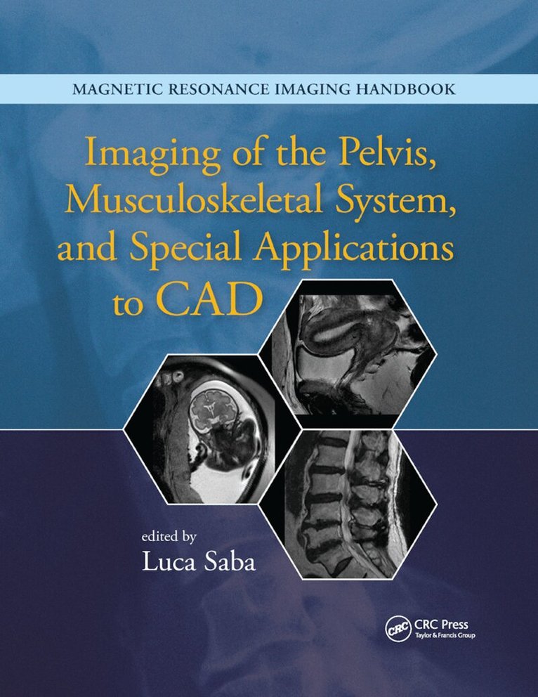
Imaging of the Pelvis, Musculoskeletal System, and Special Applications to CAD
Av Luca Saba
Häftad, 2019
1958 kr
Lägg i varukorg
Magnetic resonance imaging (MRI) is a technique used in biomedical imaging and radiology to visualize internal structures of the body. Because MRI provides excellent contrast between different soft tissues, the technique is especially useful for diagnostic imaging of the brain, muscles, and …
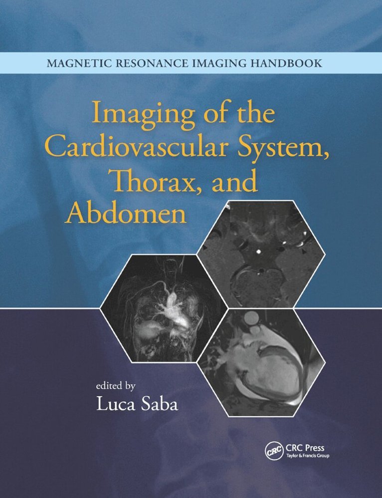
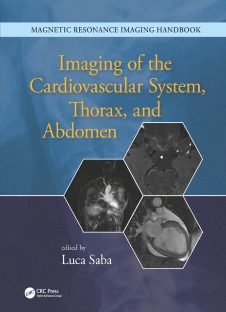
Imaging of the Cardiovascular System, Thorax, and Abdomen
Av Luca Saba
Häftad, 2019
2391 kr
Lägg i varukorg
Magnetic resonance imaging (MRI) is a technique used in biomedical imaging and radiology to visualize internal structures of the body. Because MRI provides excellent contrast between different soft tissues, the technique is especially useful for diagnostic imaging of the brain, muscles, and …
COVID-19 Radiological Lung Imaging
A Classic Artificial Intelligence Framework
Av Luca Saba, Sushant Agarwal m. fl.
Häftad, 2045
2297 kr
Kommande
COVID-19 Radiological Lung Imaging: A Classic Artificial Intelligence Framework introduces modern AI technologies for early detection of COVID-19 disease to assist in saving patient lives and safeguarding frontline workers. With a strong focus on Deep Learning, the book examines specific detection …
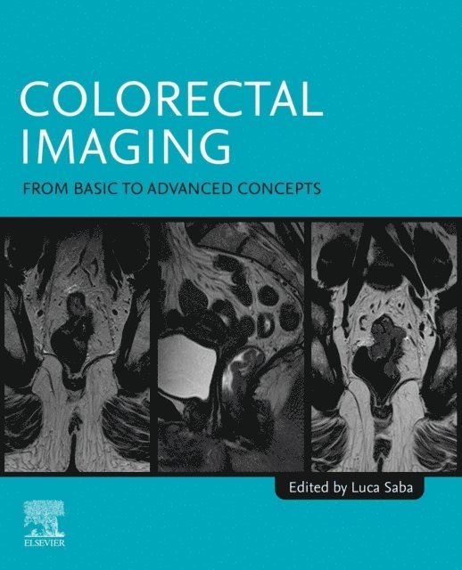
Colorectal Imaging
From Basic to Advanced Concepts
Av Luca Saba
Häftad, 2025
1740 kr
Lägg i varukorg
Colorectal Imaging: From Basic to Advanced Concepts provides an exhaustive overview of today’s basic and advanced principles of colorectal imaging, focusing not only on oncologic but also inflammatory, infective, and ischemic pathologies and complications. The book reviews basic colorectal cancer …
Colorectal Imaging
From Basic to Advanced Concepts
Av Luca Saba
E-bok, 2025
2249 kr
Lägg i varukorg
Colorectal Imaging: From Basic to Advanced Concepts provides an exhaustive overview of today''s basic and advanced principles of colorectal imaging, focusing not only on oncologic but also inflammatory, infective, and ischemic pathologies and complications. The book reviews basic colorectal cancer …
Multiple Faces of Takotsubo Syndrome
Clinical Presentations, Mechanisms and Advanced Diagnostic Insights
Inbunden, 2027
2327 kr
Kommande
Multiple Faces of Takotsubo Syndrome: Clinical Presentations, Mechanisms and Advanced Diagnostic Insights offers a thorough review of Takotsubo syndrome and the role of advanced imaging techniques in improving diagnosis, monitoring, and management of this complex cardiac condition. The book begins …
Neurobiology and Artificial Intelligence
Medical, Social and Ethical Implications
Av Luca Saba
Häftad, 2026
1991 kr
Lägg i varukorg
Neurobiology and Artificial Intelligence: Medical, Social, and Ethical Implications explores the dynamic intersection of neurobiology and artificial intelligence (AI), investigating the scientific, philosophical, and ethical implications of these rapidly evolving fields. By blending cutting-edge …
Neurobiology and Artificial Intelligence
Medical, Social and Ethical Implications
Av Luca Saba
E-bok, 2026
2575 kr
Lägg i varukorg
Neurobiology and Artificial Intelligence: Medical, Social, and Ethical Implicationsexplores the dynamic intersection of neurobiology and artificial intelligence (AI),investigating the scientific, philosophical, and ethical implications of these rapidlyevolving fields. By blending cutting-edge …
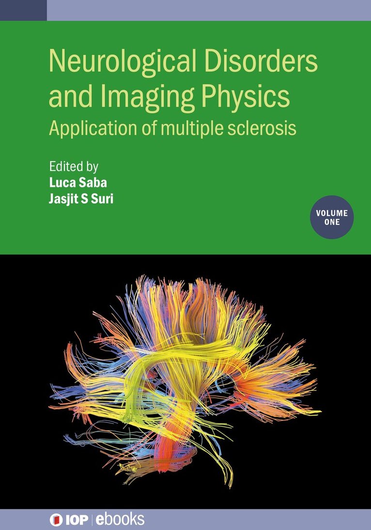
Neurological Disorders and Imaging Physics, Volume 1
Application of multiple sclerosis
Av Luca Saba, Jasjit S. Suri m. fl.
Inbunden, 2019
2029 kr
Lägg i varukorg
Multiple sclerosis (MS) is a disease where the body's immune system attacks the brain and spinal cord, causing debilitating side effects that last a lifetime for those affected. There is currently no cure. Magnetic resonance imaging (MRI) has become an established tool in the diagnosis and …
Neurological Disorders and Imaging Physics, Volume 1
Application of multiple sclerosis
Av Luca Saba, Jasjit S Suri m. fl.
Häftad, 2019
435 kr
Lägg i varukorg
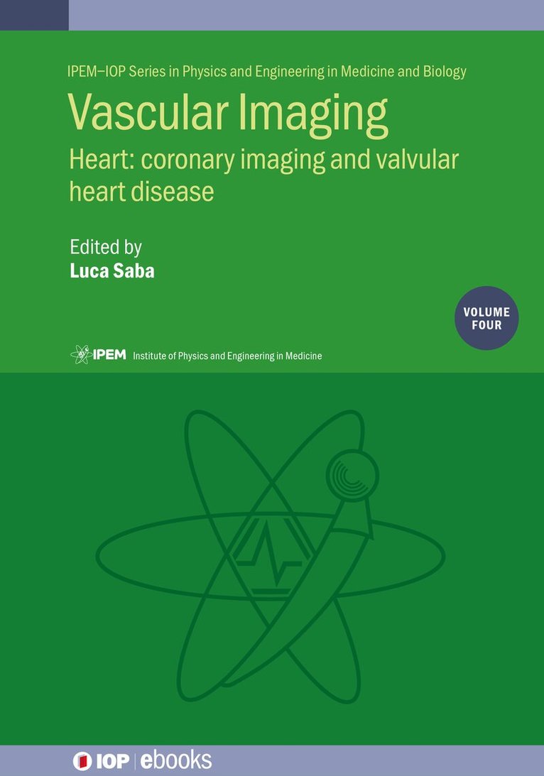
Vascular Imaging Volume 4
Heart: coronary imaging and valvular heart disease
Inbunden, 2026
1895 kr
Kommande
Magnetic Resonance Imaging Handbook, Four-Volume Set
Av Luca Saba
2027
18323 kr
Kommande
This set provides thorough coverage of medical engineering and clinical benefits in the diagnosis of human pathologies using MRI. The second edition aims to confirm itself as the leading reference for MRI application to the study of the pathological diseases of the human body including the most …
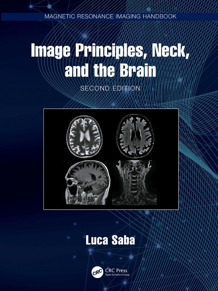
Image Principles, Neck, and the Brain
Av Luca Saba
Inbunden, 2027
3113 kr
Kommande
This book includes detailed coverage of MRI clinical applications of the head, neck and brain pathologies. Main topics are treated by leading authors in the head and neck field with outstanding quality images providing an easy usable and exhaustive text both for residents and skilled …
Imaging of Musculoskeletal System and Special Applications to CAD
Av Luca Saba
Inbunden, 2027
2788 kr
Kommande
This volume provides a comprehensive overview of the countless MRI applications in the evaluation and diagnosis of the musculoskeletal system, introducing a brand new set of chapters on CAD applications.Imaging of Musculoskeletal System and Special Applications to CAD, Second Edition, includes …
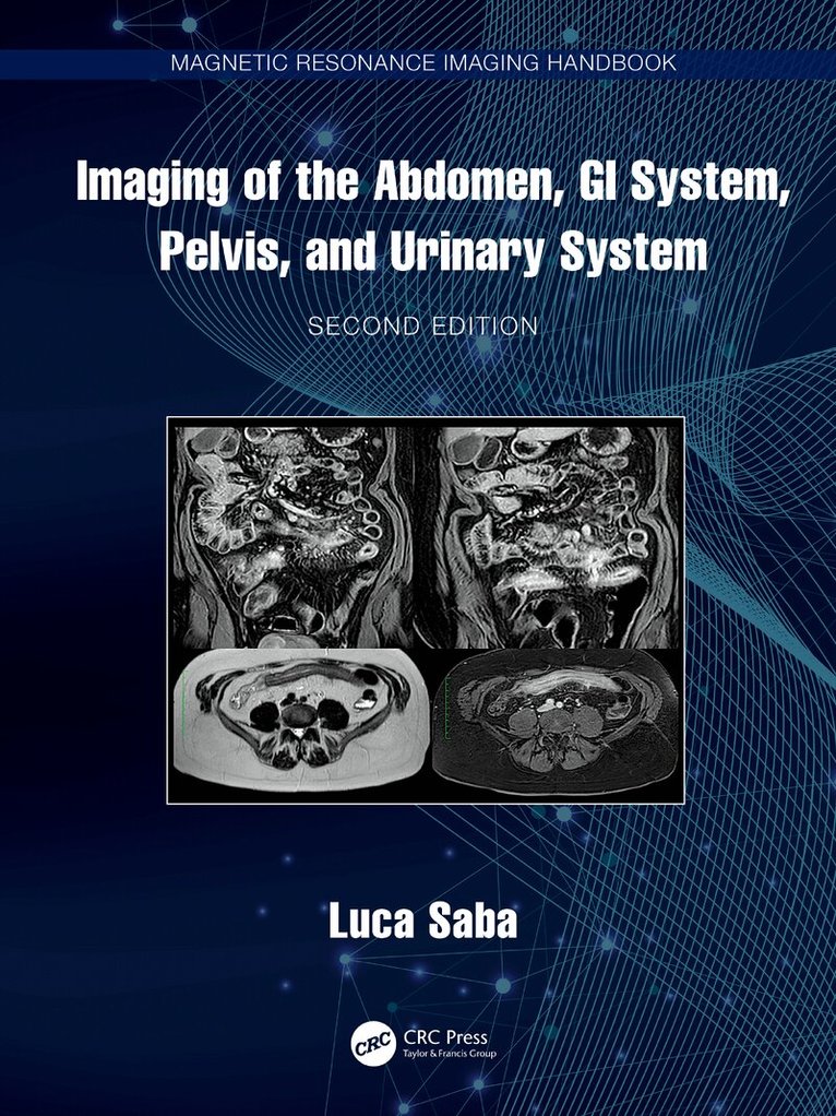
Imaging of the Abdomen, GI System, Pelvis, and Urinary System
Av Luca Saba
Inbunden, 2027
3113 kr
Kommande
This volume focuses on abdominal MRI techniques with a complete overview ranging from liver main pathologies to stomach, colon up to kidney and male and female pelvis diseases.Imaging of the Abdomen, GI System, Pelvis, and Urinary System provides a comprehensive guide to all main abdomen …
Imaging of the Cardiovascular System and Thorax
Av Luca Saba
Inbunden, 2027
2788 kr
Kommande
This book includes coverage of Cardiovascular Magnetic Resonance (CMR) applications from cardiac physiology to the interpretation and diagnosis of cardiovascular and chest diseases providing up-to-date information on CMR uses, in cardiovascular care and thoracic pathologies from basic principles to …
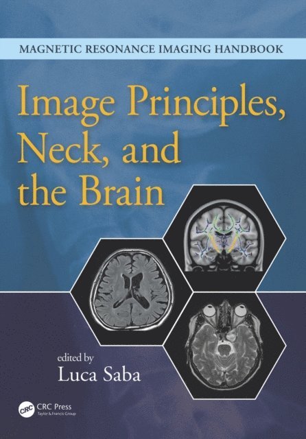
Image Principles, Neck, and the Brain
Av Luca Saba
E-bok, 2016
2251 kr
Lägg i varukorg
Magnetic resonance imaging (MRI) is a technique used in biomedical imaging and radiology to visualize internal structures of the body. Because MRI provides excellent contrast between different soft tissues, the technique is especially useful for diagnostic imaging of the brain, muscles, and …
Multi-Detector CT Imaging
Abdomen, Pelvis, and CAD Applications
E-bok, 2013
1516 kr
Lägg i varukorg
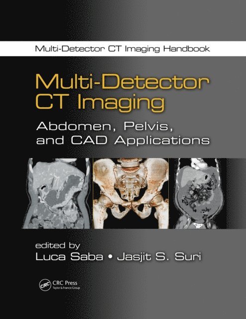
Developments in CT technology during the last 20 years have impressively improved its diagnostic potentialities. Part of a two-volume set that covers all aspects of CT imaging, Multi-Detector CT Imaging: Abdomen, Pelvis, and CAD Applications contains easily searchable clinical specialty chapters …
Multi-Detector CT Imaging
Principles, Head, Neck, and Vascular Systems
E-bok, 2013
1516 kr
Lägg i varukorg
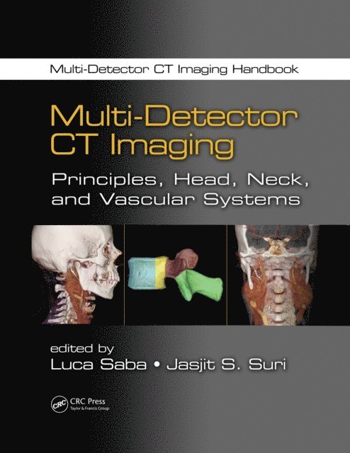
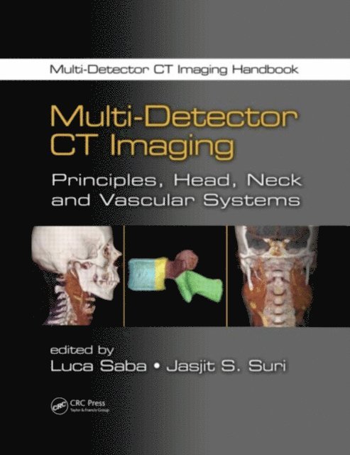
Developments in CT technology during the last 20 years have impressively improved its diagnostic potentialities. Part of a two-volume set that covers all aspects of CT imaging, Multi-Detector CT Imaging: Principles, Head, Neck, and Vascular Systems contains easily searchable clinical specialty …
Multi-Detector CT Imaging
Abdomen, Pelvis, and CAD Applications
Häftad, 2017
1282 kr
Lägg i varukorg
Developments in CT technology during the last 20 years have impressively improved its diagnostic potentialities. Part of a two-volume set that covers all aspects of CT imaging, Multi-Detector CT Imaging: Abdomen, Pelvis, and CAD Applications contains easily searchable clinical specialty chapters …
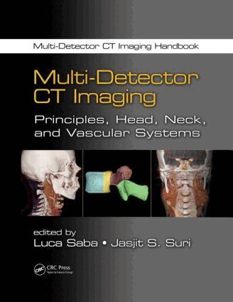
Multi-Detector CT Imaging
Principles, Head, Neck, and Vascular Systems
Häftad, 2017
1282 kr
Lägg i varukorg
Developments in CT technology during the last 20 years have impressively improved its diagnostic potentialities. Part of a two-volume set that covers all aspects of CT imaging, Multi-Detector CT Imaging: Principles, Head, Neck, and Vascular Systems contains easily searchable clinical specialty …
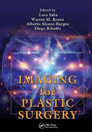
Imaging for Plastic Surgery
Av Luca Saba, Warren M. Rozen m. fl.
Häftad, 2018
1492 kr
Lägg i varukorg
Preoperative imaging is increasingly being adopted for preoperative planning in plastic and reconstructive surgery. Accurate preoperative analysis can reduce the length of operations and maximize surgical design and dissection techniques. Imaging for Plastic Surgery covers the techniques, …
Imaging of the Cardiovascular System, Thorax, and Abdomen
Av Luca Saba
E-bok, 2017
2318 kr
Lägg i varukorg
Magnetic resonance imaging (MRI) is a technique used in biomedical imaging and radiology to visualize internal structures of the body. Because MRI provides excellent contrast between different soft tissues, the technique is especially useful for diagnostic imaging of the brain, muscles, and heart. …
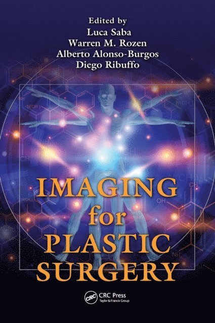
Imaging for Plastic Surgery
Av Diego Ribuffo, Alberto Alonso-Burgos m. fl.
E-bok, 2018
1765 kr
Lägg i varukorg
Preoperative imaging is increasingly being adopted for preoperative planning in plastic and reconstructive surgery. Accurate preoperative analysis can reduce the length of operations and maximize surgical design and dissection techniques. Imaging for Plastic Surgery covers the techniques, …
Multi-Detector CT Imaging
Principles, Head, Neck, and Vascular Systems
Inbunden, 2013
3533 kr
Lägg i varukorg
Developments in CT technology during the last 20 years have impressively improved its diagnostic potentialities. Part of a two-volume set that covers all aspects of CT imaging, Multi-Detector CT Imaging: Principles, Head, Neck, and Vascular Systems contains easily searchable clinical specialty …
Multi-Detector CT Imaging
Principles, Head, Neck, and Vascular Systems
E-bok, 2013
1516 kr
Lägg i varukorg
Developments in CT technology during the last 20 years have impressively improved its diagnostic potentialities. Part of a two-volume set that covers all aspects of CT imaging, Multi-Detector CT Imaging: Principles, Head, Neck, and Vascular Systems contains easily searchable clinical specialty …