• Fri frakt över 249 kr
  • •
  • Snabba leveranser
  • •
  • Billiga böcker
Kundservice

Du är på sajten för privatpersoner.

Företag, bibliotek eller offentlig verksamhet?

Du handlar på classic.bokus.com, där alla dina funktioner finns intakta.
Till classic.bokus.com
Bokus logotyp. Gå till startsidan.
  • Erbjudanden
  • Nyheter
  • Student
  • Topplistor
  • Barn & ungdom
  • Bokus Play
  • E-böcker
  • Pocketböcker
  • Spel & pussel

10% rabatt på allt med kod: NYSTART10 →

Sidfot

Mina sidor

    Hjälp

    • Kundservice
    • Vanliga frågor och svar
    • Frakt och leverans
    • Retur vid ångerrätt
    • Reklamera vara
    • Betalning
    • Köpvillkor
    • Allmänna villkor
    • Information om webbplatsens tillgänglighet

    Om Bokus

    • Om oss
    • Pressrum
    • För studenter
    • För företag
    • För bibliotek och offentlig verksamhet
    • För leverantörer
    • Hållbarhet

    Populärt

    • Aktuella erbjudanden
    • Presentkort
    • Studentlitteratur
    • Nya böcker
    • Topplistor
    • Signerade böcker
    • Engelska böcker

    Inspiration

    • Boktips
    • BookTok
    • Populära bokserier
    • Barnbokskaraktärer
    • Populära författare
    Logotyp för Bokus
    Följ oss på Facebook (extern länk)Följ oss på Instagram (extern länk)Följ oss på YouTube (extern länk)Följ oss på TikTok (extern länk)
    bokus @ CookiesAnpassa cookiesIntegritetspolicyKöpvillkor
    Till Citymail hemsida (extern länk)Till Budbee hemsida (extern länk)Till Postnord hemsida (extern länk)Till Schenker hemsida (extern länk)Till Early Bird hemsida (extern länk)Till Walleys hemsida (extern länk)
    1. Medicin
    2. Klinisk medicin och internmedicin
    3. Sjukdomar och rubbningar
    4. Onkologi

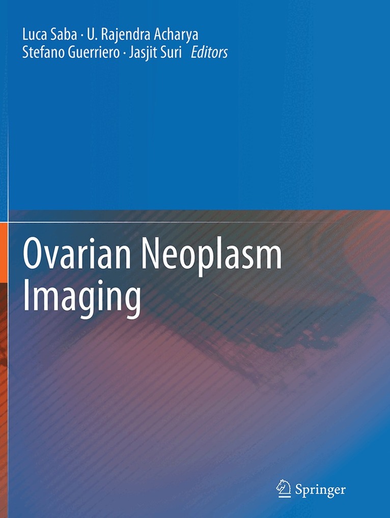
    Ovarian Neoplasm Imaging

    AvLuca Saba,U. Rajendra Acharya

    Häftad, Engelska, 2016

    2 229 kr

    Beställningsvara. Skickas inom 10-15 vardagar. Fri frakt över 249 kr.

    Beskrivning

    ​Diagnostic and pre-operative imaging has become increasingly adopted throughout the field of gynecology. In particular accurate preoperative analysis of ovarian pathology can improve the selection of the correct therapeutical approach, to reduce the length of operations, maximize surgical technique, and can ultimately improve a range of operative outcomes. New imaging modalities have advanced to the point of high resolution, three-dimensional analysis of tissue anatomy, composition and perfusion. Ultrasonography (US), Computed tomographic (CT) and magnetic resonance (MR) imaging have recently emerged as outstanding non-invasive techniques for the detection and characterization of ovarian pathology. In particular, US has probably now imposed itself as the “state-of-the-art” technique to explore the ovarian neoplasm, thanks the technical advancement like texture analysis (MGV) and 3D potentialities, although MR with new sequences like the diffusion-weighted imaging and greater magnetic fields (3 Tesla or more) may become leading methods in the future. The purpose of this book is to cover all the imaging techniques, potential for applying such imaging clinically, and to offer present and future applications as applied to ovarian pathology with the most world renowned scientists in these fields. The book is designed according to the pathological classification of the benign and malignant ovarian neoplasm by presenting for each pathology the clinical setting followed by the imaging approach. At the end of each chapter a “focus concept” paragraph will be presented with take-home point for the readers.

    Produktinformation

    • Utgivningsdatum:2016-09-29
    • Mått:210 x 279 x 28 mm
    • Vikt:1 469 g
    • Format:Häftad
    • Språk:Engelska
    • Antal sidor:535
    • Förlag:Springer-Verlag New York Inc.
    • ISBN:9781489979513

    Utforska kategorier

    • Onkologi inom Medicin
    • Medicinsk bildbehandling inom Medicin
    • Gynekologi och obstetrik inom Medicin

    Mer om författaren

    Luca Saba received the MD from the University of Cagliari, Italy in 2002. Today he works in the A.O.U. of Cagliari. He is member of the Italian Society of Radiology (SIRM), European Society of Radiology (ESR), Radiological Society of North America (RSNA), American Roentgen Ray Society (ARRS) and European Society of Neuroradiology (ESNR). Rajendra Acharya, PhD, DEng is a Visiting faculty in Ngee Ann Polytechnic, Singapore, Adjunct faculty in Singapore Institute of Technology- University of Glasgow degree programme, Singapore, Associate faculty in SIM University, Singapore and Adjunct faculty in Manipal Institute of Technology, Manipal, India. He received his Ph.D. from National Institute of Technology Karnataka, Surathkal, India and D Engg from Chiba University, Japan. Stefano Guerriero MD, Born Siracusa (Italy) 10 October 1961. Medical doctor University of Pisa 24 October 1988. Postgraduate in Obstetrics and Gynecology University of Pisa october 1992. He is Associate Professor of obstetrics and gynecology at The University of Cagliari. Editor of Ultrasound in Obstetrics and Gynecology from 2011 Jasjit S. Suri, MS, PhD, MBA, received his Masters from University of Illinois, Chicago, Doctorate from University of Washington, Seattle, and Executive Management from Weatherhead School of Management, Case Western Reserve University (CWRU), Cleveland. Dr. Suri was crowned with President’s Gold medal in 1980 and the Fellow of American Institute of Medical and Biological Engineering (AIMBE) for his outstanding contributions at Washington DC.

    Innehållsförteckning

    • Epidemiology.- Histopathology.- Cyst of Follicular origin and Pregnancy Luteoma(CT and MR).-Endometrioma (Clinical Setting & US).- Endometrioma (CT and MR).- Benign Surface.- Epithelial Stromal Tumors (Clinical Setting & US).- Benign Surface Epithelial Stromal Tumors (CT and MR).- Benign Sex Cord - Stromal Tumors (Clinical Setting & US).- BenignSex Cord - Stromal Tumors (CT and MR).- Benign Germ Cell - Stromal Tumors (Clinical Setting & US).- Benign Germ Cell - Stromal Tumors (CT and MR).- Borderline Tumor (Serous\Mucinous\Endometrioid) (Clinical Setting & US).- Borderline Tumor (Serous\Mucinous\Endometrioid) (CT and MR).- Malignant Tumor (Serous\Mucinous\Endometrioid adenocarcinoma) (Clinical Setting & US).- Malignant Tumor (Serous\Mucinous\Endometrioid adenocarcinoma) (CT and MR).- Rare Malignant Tumor (Clear cell adenocarcinoma, transitional cell carcinoma, malignant Brenner Tumor) (Clinical Setting & US).- Rare Tumor (Clear cell adenocarcinoma, transitional cell carcinoma, malignant Brenner Tumor) (CT and MR).- Malignant Sex Cord - Stromal Tumors (Clinical Setting & US).- Malignant Sex Cord - Stromal Tumors (CT and MR).- Malignant Germ Cell - Stromal Tumors (Clinical Setting & US).- Malignant Germ Cell - Stromal Tumors (CT and MR).- Metastatic tumors (Clinical Setting & US).- Metastatic tumors (Serous\Mucinous\Endometrioid) (CT and MR).- 3D Ultrasonography.- Ovarian Tumor Characterization and classification using ultrasound : A new on-line paradigm.- Ovarian Tumor Characterization using 3D ultrasound.- Evolutionary Algorithm based Classifier Parameter Tuning for automatic Ovarian Cancer tissue characteization and Classification.- CT/PET.- Contrast-enhanced transvaginal sonography for early detection of ovarian cancer.- Molecular imaging.- Index.