Ayman El-Baz – författare
Visar alla böcker från författaren Ayman El-Baz. Handla med fri frakt och snabb leverans.
85 produkter
Handbook of Texture Analysis, Two-Volume Set
Two-Volume Set
Av Ayman El-Baz, Ghazal Mohammad A. m. fl.
2024
5878 kr
Lägg i varukorg
This two-volume handbook provides a comprehensive view of texture analysis in both AI-based industrial and medical imaging applications. The first volume covers texture analysis for neuroradiology; information-theoretic entropy; chronic liver diseases; clinical management of focal liver lesions; …
Handbook of Texture Analysis
Generalized Texture for AI-Based Industrial Applications
Av Ayman El-Baz, Mohammad Ghazal m. fl.
Inbunden, 2024
2601 kr
Lägg i varukorg
The major goals of texture research in computer vision are to understand, model, and process texture, and ultimately, to simulate the human visual learning process using computer technologies. In the last decade, artificial intelligence has been revolutionized by machine learning and big data …
Handbook of Texture Analysis
AI-Based Medical Imaging Applications
Av Ayman El-Baz, Mohammed Ghazal m. fl.
Inbunden, 2024
2601 kr
Lägg i varukorg
The major goals of texture research in computer vision are to understand, model, and process texture and, ultimately, to simulate the human visual learning process using computer technologies. In the last decade, artificial intelligence has been revolutionized by machine learning and big data …
Cloud Computing in Medical Imaging
Av Ayman El-Baz, Jasjit S. Suri
Inbunden, 2023
2601 kr
Lägg i varukorg
Today’s healthcare organizations must focus on a lot more than just the health of their clients. The infrastructure it takes to support clinical-care delivery continues to expand, with information technology being one of the most significant contributors to that growth. As companies have become …
Biomedical Image Segmentation
Advances and Trends
Av Ayman El-Baz, Xiaoyi Jiang m. fl.
Häftad, 2019
1968 kr
Lägg i varukorg
As one of the most important tasks in biomedical imaging, image segmentation provides the foundation for quantitative reasoning and diagnostic techniques. A large variety of different imaging techniques, each with its own physical principle and characteristics (e.g., noise modeling), often requires …
Autism Imaging and Devices
Av Manuel F. Casanova, Ayman El-Baz m. fl.
Häftad, 2019
1229 kr
Lägg i varukorg
This book covers state-of-the-art medical image analysis approaches currently pursued in autism research. Chapters cover recent advances in diagnosis using structural neuroimaging. All aspects of imaging are included, such as electrophysiology (EEG, ERP, QEEG, and MEG), postmortem techniques, and …
Lung Imaging and CADx
E-bok, 2019
999 kr
Lägg i varukorg
Developing an effective computer-aided diagnosis (CAD) system for lung cancer is of great clinical importance and can significantly increase the patient''s chance for survival. For this reason, CAD systems for lung cancer have been investigated in a large number of research studies. A typical CAD …
Lung Imaging and CADx
E-bok, 2019
999 kr
Lägg i varukorg
Developing an effective computer-aided diagnosis (CAD) system for lung cancer is of great clinical importance and can significantly increase the patient''s chance for survival. For this reason, CAD systems for lung cancer have been investigated in a large number of research studies. A typical CAD …
Machine Learning in Medicine
Av Jasjit S. Suri, Ayman El-Baz
E-bok, 2021
1480 kr
Lägg i varukorg
Machine Learning in Medicine covers the state-of-the-art techniques of machine learning and their applications in the medical field. It presents several computer-aided diagnosis (CAD) systems, which have played an important role in the diagnosis of several diseases in the past decade, e.g., cancer …
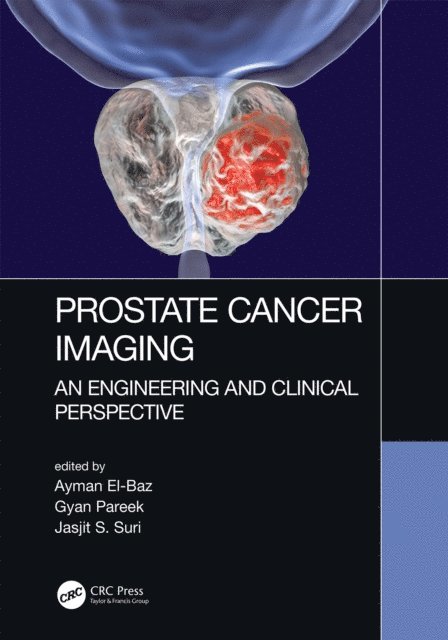
Prostate Cancer Imaging
An Engineering and Clinical Perspective
Av Jasjit S. Suri, Gyan Pareek m. fl.
E-bok, 2018
977 kr
Lägg i varukorg
This book covers novel strategies and state of the art approaches for automated non-invasive systems for early prostate cancer diagnosis. Prostate cancer is the most frequently diagnosed malignancy after skin cancer and the second leading cause of cancer related male deaths in the USA after lung …
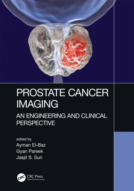
Prostate Cancer Imaging
An Engineering and Clinical Perspective
Av Jasjit S. Suri, Gyan Pareek m. fl.
E-bok, 2018
977 kr
Lägg i varukorg
This book covers novel strategies and state of the art approaches for automated non-invasive systems for early prostate cancer diagnosis. Prostate cancer is the most frequently diagnosed malignancy after skin cancer and the second leading cause of cancer related male deaths in the USA after lung …
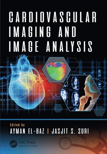
Cardiovascular Imaging and Image Analysis
Av Jasjit S. Suri, Ayman El-Baz
E-bok, 2018
4449 kr
Lägg i varukorg
This book covers the state-of-the-art approaches for automated non-invasive systems for early cardiovascular disease diagnosis. It includes several prominent imaging modalities such as MRI, CT, and PET technologies. There is a special emphasis placed on automated imaging analysis techniques, which …
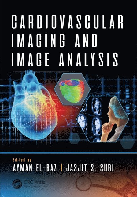
Cardiovascular Imaging and Image Analysis
Av Jasjit S. Suri, Ayman El-Baz
E-bok, 2018
4547 kr
Lägg i varukorg
This book covers the state-of-the-art approaches for automated non-invasive systems for early cardiovascular disease diagnosis. It includes several prominent imaging modalities such as MRI, CT, and PET technologies. There is a special emphasis placed on automated imaging analysis techniques, which …
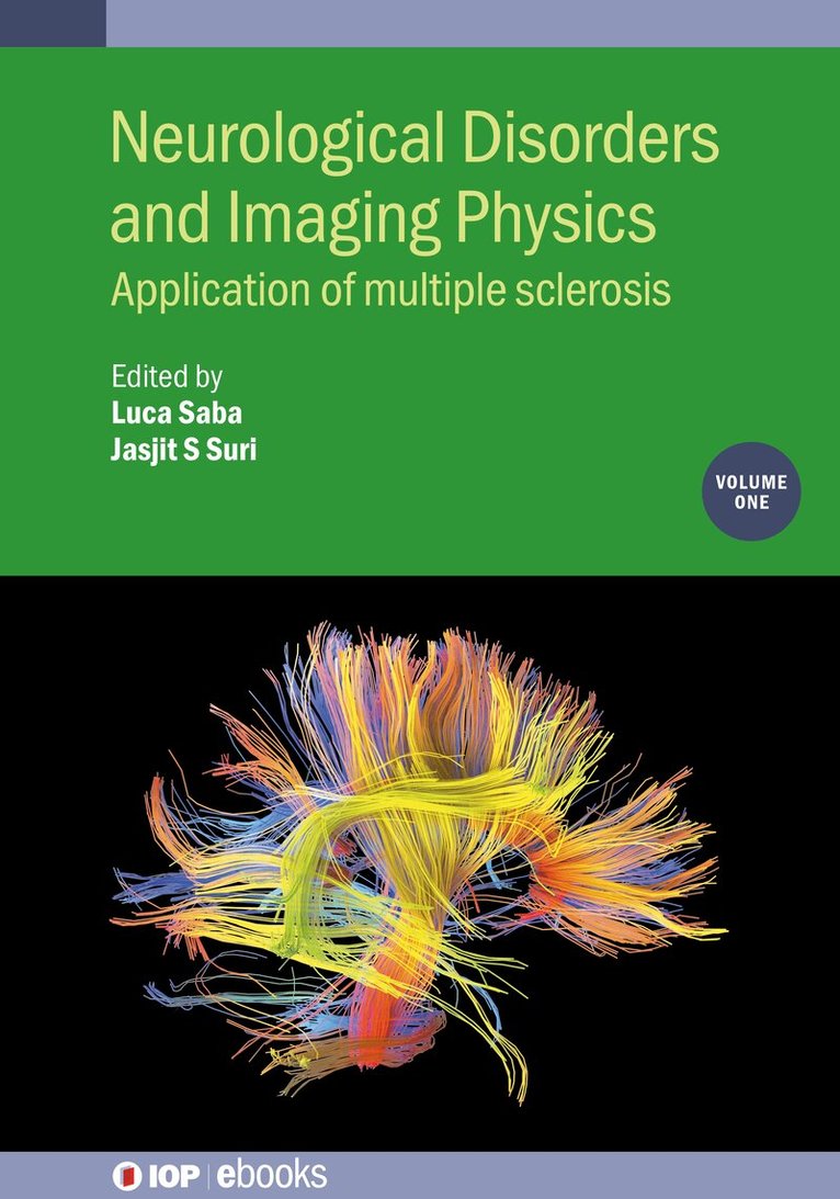
Neurological Disorders and Imaging Physics, Volume 1
Application of multiple sclerosis
Av Luca Saba, Jasjit S. Suri m. fl.
Inbunden, 2019
2029 kr
Lägg i varukorg
Multiple sclerosis (MS) is a disease where the body's immune system attacks the brain and spinal cord, causing debilitating side effects that last a lifetime for those affected. There is currently no cure. Magnetic resonance imaging (MRI) has become an established tool in the diagnosis and …
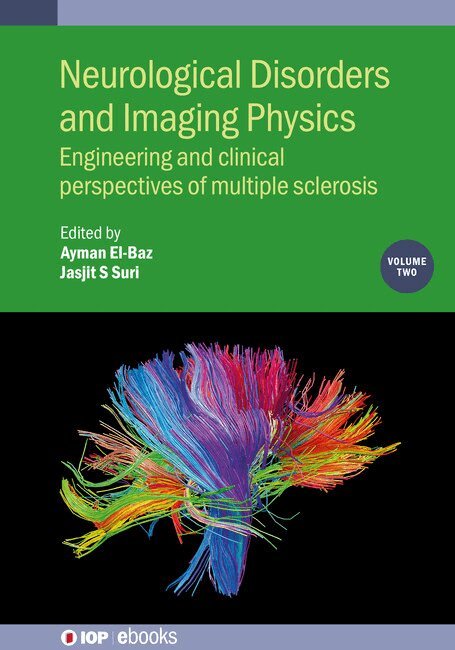
Neurological Disorders and Imaging Physics, Volume 2
Engineering and clinical perspectives of multiple sclerosis
Av Ayman El-Baz, Jasjit S. Suri m. fl.
Inbunden, 2019
2029 kr
Lägg i varukorg
More than two million people worldwide were affected by multiple sclerosis (MS) in 2015, with rates varying widely among different populations. This book explores the basics of MS, which is the most common immune-mediated disorder affecting the central nervous system. Multiple sclerosis is a …
Neurological Disorders and Imaging Physics, Volume 2
Engineering and clinical perspectives of multiple sclerosis
Av Ayman El-Baz, Jasjit S Suri
Häftad, 2019
426 kr
Lägg i varukorg
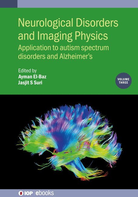
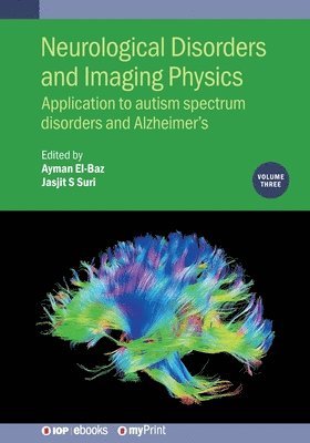
Neurological Disorders and Imaging Physics, Volume 3
Application to autism spectrum disorders and Alzheimer’s
Av Ayman El-Baz, Jasjit S. Suri m. fl.
Inbunden, 2019
2029 kr
Lägg i varukorg
Autism spectrum disorder (ASD) and Alzheimer's disease (AD) are two significant neurological disorders, which represent the scope of this book. Both ASD and AD affect a significant number of the population and present in numerous ways. This volume covers the state-of-the-art topics that investigate …
Neurological Disorders and Imaging Physics, Volume 3
Application to autism spectrum disorders and Alzheimer's
Av Ayman El-Baz, Jasjit S Suri
Häftad, 2019
426 kr
Lägg i varukorg
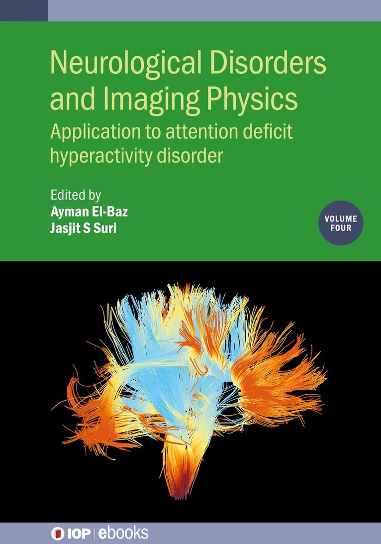
Neurological Disorders and Imaging Physics, Volume 4
Application to attention deficit hyperactivity disorder
Av Ayman El-Baz, Jasjit S Suri
Inbunden, 2020
2029 kr
Lägg i varukorg
Neurological Disorders and Imaging Physics, Volume 4
Application to attention deficit hyperactivity disorder
Av Ayman El-Baz, Ayman El-Baz m. fl.
Häftad, 2020
435 kr
Lägg i varukorg
Neurological Disorders and Imaging Physics, Volume 1
Application of multiple sclerosis
Av Luca Saba, Jasjit S Suri m. fl.
Häftad, 2019
435 kr
Lägg i varukorg
Photo Acoustic and Optical Coherence Tomography Imaging, Volume 1
Diabetic retinopathy
Av Ayman El-Baz, Jasjit S. Suri m. fl.
Inbunden, 2023
2783 kr
Lägg i varukorg
Photo Acoustic and Optical Coherence Tomography Imaging, Volume 1
Diabetic retinopathy
Av Ayman El-Baz, Jasjit S Suri
Häftad, 2023
509 kr
Lägg i varukorg
Photo Acoustic and Optical Coherence Tomography Imaging, Volume 2
Fundus imaging for the retina
Av Ayman El-Baz, Jasjit S. Suri m. fl.
Inbunden, 2022
2783 kr
Lägg i varukorg
Photo Acoustic and Optical Coherence Tomography Imaging, Volume 2
Fundus imaging for the retina
Av Ayman El-Baz, Jasjit S Suri
Häftad, 2022
509 kr
Lägg i varukorg
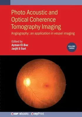
Photo Acoustic and Optical Coherence Tomography Imaging, Volume 3
Angiography: an application in vessel imaging
Av Ayman El-Baz, Jasjit S. Suri m. fl.
Inbunden, 2021
1985 kr
Lägg i varukorg
This book covers the state-of-the-art techniques of optical coherence tomography angiography (OCTA) imaging for the diagnosis of retinal diseases. It is part of a three-volume work that describes the latest imaging techniques in which to bring optical coherence tomography (OCT), Fundus Imaging and …
Photo Acoustic and Optical Coherence Tomography Imaging, Volume 3
Angiography: an application in vessel imaging
Av Ayman El-Baz, Jasjit S Suri
Häftad, 2021
435 kr
Lägg i varukorg
Lung Cancer and Imaging
Av Ayman El-Baz, Jasjit S. Suri m. fl.
Inbunden, 2019
2029 kr
Lägg i varukorg
Lung cancer is one of the most common cancers in men and women worldwide. Early diagnosis of lung cancer can significantly increase the chances of a patient's survival, yet early detection has historically been difficult. As a result, there has been a great deal of progress in the development of …
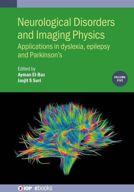
Neurological Disorders and Imaging Physics, Volume 5
Applications in dyslexia, epilepsy and Parkinson’s
Av Jasjit S Suri, Ayman El-Baz
Inbunden, 2020
2029 kr
Lägg i varukorg
This book focuses on major trends and challenges inthe area of dyslexia, epilepsy and Parkinson's, and aims to identify newtechniques and their applications in biomedical analysis. This is an essentialreference for students and researchers in medical imaging, brain imaging, imageprocessing, and …