Mike Leach – författare
1 554 kr
Skickas inom 5-8 vardagar
1 542 kr
Skickas inom 5-8 vardagar
1 808 kr
Läs direkt efter köp
1 745 kr
Läs direkt efter köp
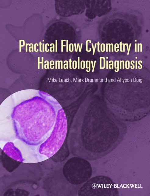
The accurate diagnosis of haematologic malignancies is a complex and challenging task. It routinely involves morphologic, molecular, cytogenetic and flow cytometric expertise. To determine what treatment protocol will be followed, it is vital to integrate, interpret and report these results accurately.
Flow cytometry is key in this diagnostic pathway.
This book guides the reader as to how flow cytometry results should be interpreted and applied to optimize patient care. At the core of this text is an appreciation of clinical, morphological and immunophenotypic correlation and the importance of constant liaison and discussion between the medical and scientific teams.
The authors present a logical and practical approach to the diagnosis of blood disorders (both neoplastic and reactive) and evaluate the diagnostic applications of flow cytometry.
Practical Flow Cytometry in Haematology Diagnosis provides:
A clinical reference source on all aspects of flow cytometry, covering both malignant and benign conditionsCarefully chosen real-life cases in each chapter, complemented by high quality morphological images Help in making a diagnosis, together with an understanding of the limitations of the technique and the potential pitfallsAll those who instigate, perform, interpret or act upon flow cytometry patient material will find this book an invaluable guide.
1 760 kr
Läs direkt efter köp
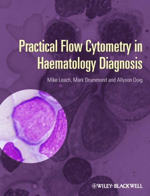
The accurate diagnosis of haematologic malignancies is a complex and challenging task. It routinely involves morphologic, molecular, cytogenetic and flow cytometric expertise. To determine what treatment protocol will be followed, it is vital to integrate, interpret and report these results accurately.
Flow cytometry is key in this diagnostic pathway.
This book guides the reader as to how flow cytometry results should be interpreted and applied to optimize patient care. At the core of this text is an appreciation of clinical, morphological and immunophenotypic correlation and the importance of constant liaison and discussion between the medical and scientific teams.
The authors present a logical and practical approach to the diagnosis of blood disorders (both neoplastic and reactive) and evaluate the diagnostic applications of flow cytometry.
Practical Flow Cytometry in Haematology Diagnosis provides:
A clinical reference source on all aspects of flow cytometry, covering both malignant and benign conditionsCarefully chosen real-life cases in each chapter, complemented by high quality morphological images Help in making a diagnosis, together with an understanding of the limitations of the technique and the potential pitfallsAll those who instigate, perform, interpret or act upon flow cytometry patient material will find this book an invaluable guide.
1 572 kr
Läs direkt efter köp
The analysis of blood, bone marrow and tissue fluid specimens requires a multi-faceted approach with the integration of scientific data from a number of disciplines. No single discipline can operate in isolation or errors will occur. Flow cytometry is in a privileged position in that it can provide rapid analysis of specimens and it is often the first definitive investigation to produce results and help formulate a working diagnosis.
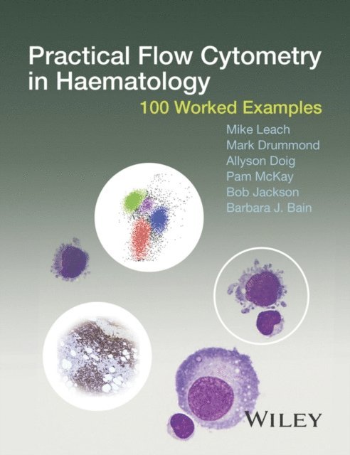
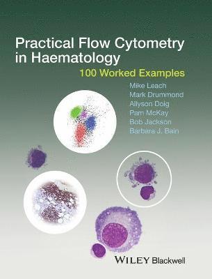
This companion text to Practical Flow Cytometry in Haematology Diagnosis contains 100 worked examples drawn from real clinical cases presenting to the authors’ institution. Cases are illustrated with peripheral blood and bone marrow cytology, tissue pathology and cytogenetic and molecular data, which are integrated to generate, where appropriate, a diagnosis based on the WHO Classification of Tumours of Haematopoietic and Lymphoid Tissues. The spectrum of clinical cases includes adult and paediatric patients, and both neoplastic and reactive disorders. The cases appear in no particular order to challenge the reader to make their own diagnosis.
The reader will review May−Grünwald−Giemsa (MGG)-stained films of peripheral blood and bone marrow aspirates presented alongside flow cytometric data and haematoxylin and eosin (H&E)-stained bone marrow and other tissue biopsy sections. Immunohistochemistry is used to further clarify the tissue lineage and cell differentiation. Cytogenetic studies using metaphase preparations are used to identify translocations and chromosome gains and losses whilst interphase fluorescence in situ hybridisation (FISH) studies and polymerase chain reaction (PCR) are used to identify gene fusions, gene rearrangements and deletions. Each case concludes with a discussion of the features that are important to making a diagnosis. The cases are also listed according to disease classification in the appendix so that the text can also be used as a reference.
Practical Flow Cytometry in Haematology: 100 Worked Examples:
Provides a practical, example-based resource for flow cytometry Demonstrates how flow cytometry results should be interpreted and applied to optimize patient care Includes both malignant and benign conditions Can be used in conjunction with Practical Flow Cytometry in Haematology Diagnosis, by the same author team (ISBN 9780470671207)Practical Flow Cytometry in Haematology: 100 Worked Examples is ideal for practicing haematologists and histopathologists with an interest in haematopathology, but particularly directed at trainee haematologists and scientists preparing for FRCPath and related examinations.
1 629 kr
Läs direkt efter köp
The analysis of blood, bone marrow and tissue fluid specimens requires a multi-faceted approach with the integration of scientific data from a number of disciplines. No single discipline can operate in isolation or errors will occur. Flow cytometry is in a privileged position in that it can provide rapid analysis of specimens and it is often the first definitive investigation to produce results and help formulate a working diagnosis.
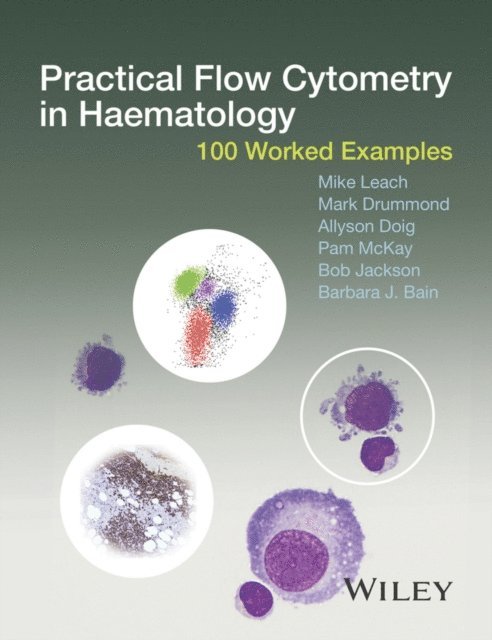
This companion text to Practical Flow Cytometry in Haematology Diagnosis contains 100 worked examples drawn from real clinical cases presenting to the authors’ institution. Cases are illustrated with peripheral blood and bone marrow cytology, tissue pathology and cytogenetic and molecular data, which are integrated to generate, where appropriate, a diagnosis based on the WHO Classification of Tumours of Haematopoietic and Lymphoid Tissues. The spectrum of clinical cases includes adult and paediatric patients, and both neoplastic and reactive disorders. The cases appear in no particular order to challenge the reader to make their own diagnosis.
The reader will review May−Grünwald−Giemsa (MGG)-stained films of peripheral blood and bone marrow aspirates presented alongside flow cytometric data and haematoxylin and eosin (H&E)-stained bone marrow and other tissue biopsy sections. Immunohistochemistry is used to further clarify the tissue lineage and cell differentiation. Cytogenetic studies using metaphase preparations are used to identify translocations and chromosome gains and losses whilst interphase fluorescence in situ hybridisation (FISH) studies and polymerase chain reaction (PCR) are used to identify gene fusions, gene rearrangements and deletions. Each case concludes with a discussion of the features that are important to making a diagnosis. The cases are also listed according to disease classification in the appendix so that the text can also be used as a reference.
Practical Flow Cytometry in Haematology: 100 Worked Examples:
Provides a practical, example-based resource for flow cytometry Demonstrates how flow cytometry results should be interpreted and applied to optimize patient care Includes both malignant and benign conditions Can be used in conjunction with Practical Flow Cytometry in Haematology Diagnosis, by the same author team (ISBN 9780470671207)Practical Flow Cytometry in Haematology: 100 Worked Examples is ideal for practicing haematologists and histopathologists with an interest in haematopathology, but particularly directed at trainee haematologists and scientists preparing for FRCPath and related examinations.
1 409 kr
Skickas inom 5-8 vardagar
1 423 kr
Skickas inom 5-8 vardagar
1 645 kr
Läs direkt efter köp
Offers clear and concise instruction on running, reporting and interpreting immunophenotyping studies
Written by two well-known haematology educators and experts on the topic, Immunophenotyping for Haematologists contains an introduction to running, reporting and interpreting immunophenotyping studies. The book offers a unique approach to the topic by putting the focus on clinical and laboratory haematologists who are not routinely involved in running and reporting on immunophenotyping studies.
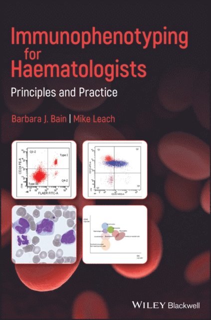
Immunophenotyping using flow cytometry has become the method of choice in identifying and sorting cells within complex populations, for example, the analysis of immune or neoplastic cells in a blood sample. The text reviews the purpose and principles of immunophenotyping and includes an introduction and explanation of the principles and the role of immunophenotyping. The authors examine immunophenotypic characteristics of the disease groups commonly encountered and identify the features that differentiate malignant cells from normal cells. To enhance understanding, the book contains multiple choice and extended matching questions which integrates immunophenotyping with clinicopathological features and the results of other investigations to mimic everyday practice. This important book:
Provides a concise introduction to running, reporting and interpreting immunophenotyping studies Contains a list of all the antibody specificities currently widely used in diagnosis and disease monitoring Presents an ideal reference for use in laboratories, including immunophenotyping laboratories Aids in the interpretation by covering immunophenotypic characteristics of commonly encountered disease groups Identifies the features that differentiate malignant cells from their normal counterpartsWritten for haematologists working in both laboratory and clinical haematology, Immunophenotyping for Haematologists is a much-needed reference for understanding and interpreting immunophenotyping studies.
1 602 kr
Läs direkt efter köp
Offers clear and concise instruction on running, reporting and interpreting immunophenotyping studies
Written by two well-known haematology educators and experts on the topic, Immunophenotyping for Haematologists contains an introduction to running, reporting and interpreting immunophenotyping studies. The book offers a unique approach to the topic by putting the focus on clinical and laboratory haematologists who are not routinely involved in running and reporting on immunophenotyping studies.
Immunophenotyping using flow cytometry has become the method of choice in identifying and sorting cells within complex populations, for example, the analysis of immune or neoplastic cells in a blood sample. The text reviews the purpose and principles of immunophenotyping and includes an introduction and explanation of the principles and the role of immunophenotyping. The authors examine immunophenotypic characteristics of the disease groups commonly encountered and identify the features that differentiate malignant cells from normal cells. To enhance understanding, the book contains multiple choice and extended matching questions which integrates immunophenotyping with clinicopathological features and the results of other investigations to mimic everyday practice. This important book:
Provides a concise introduction to running, reporting and interpreting immunophenotyping studies Contains a list of all the antibody specificities currently widely used in diagnosis and disease monitoring Presents an ideal reference for use in laboratories, including immunophenotyping laboratories Aids in the interpretation by covering immunophenotypic characteristics of commonly encountered disease groups Identifies the features that differentiate malignant cells from their normal counterpartsWritten for haematologists working in both laboratory and clinical haematology, Immunophenotyping for Haematologists is a much-needed reference for understanding and interpreting immunophenotyping studies.
1 369 kr
Skickas
1 662 kr
Läs direkt efter köp
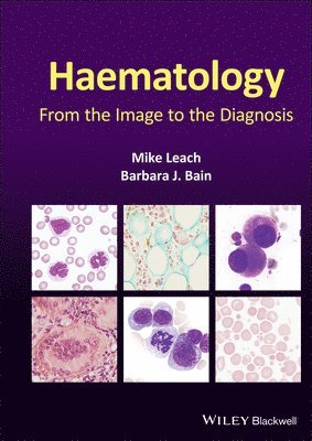
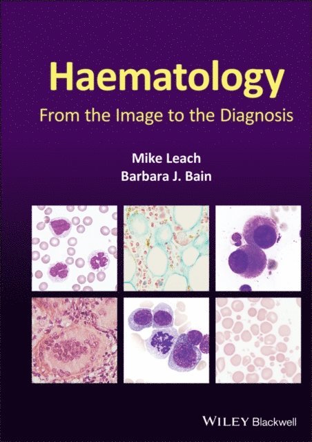
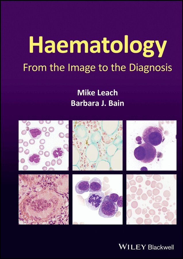
Diagnostic haematology requires the assessment of clinical and laboratory data together with a careful morphological assessment of cells in blood, bone marrow and tissue fluids. Subsequent investigations including flow cytometry, immunohistochemistry, cytogenetics and molecular studies are guided by the original morphological findings. These targeted investigations help generate a prompt unifying diagnosis. Haematology: From the Image to the Diagnosis presents a series of cases illustrating how skills in morphology can guide the investigative process. In this book, the authors capture a series of images to illustrate key features to recognize when undertaking a morphological review and show how they can be integrated with supplementary information to reach a final diagnosis.
Using a novel format of visual case studies, this text mimics ‘real life’ for the practising diagnostic haematologist – using brief clinical details and initial microscopic morphological triage to formulate a differential diagnosis and a plan for efficient and economical confirmatory investigation to deduce the correct final diagnosis. The carefully selected, high-quality photomicrographs and the clear, succinct descriptions of key features, investigations and results will help haematologists, clinical scientists, haematology trainees and haematopathologists to make accurate diagnoses in their day-to-day work.
Covering a wide range of topics, and including paediatric as well as adult cases, Haematology: From the Image to the Diagnosis is a succinct visual guide which will be welcomed by consultants, trainees and scientists alike.
1 662 kr
Läs direkt efter köp
Diagnostic haematology requires the assessment of clinical and laboratory data together with a careful morphological assessment of cells in blood, bone marrow and tissue fluids. Subsequent investigations including flow cytometry, immunohistochemistry, cytogenetics and molecular studies are guided by the original morphological findings. These targeted investigations help generate a prompt unifying diagnosis. Haematology: From the Image to the Diagnosis presents a series of cases illustrating how skills in morphology can guide the investigative process. In this book, the authors capture a series of images to illustrate key features to recognize when undertaking a morphological review and show how they can be integrated with supplementary information to reach a final diagnosis.
Using a novel format of visual case studies, this text mimics ‘real life’ for the practising diagnostic haematologist – using brief clinical details and initial microscopic morphological triage to formulate a differential diagnosis and a plan for efficient and economical confirmatory investigation to deduce the correct final diagnosis. The carefully selected, high-quality photomicrographs and the clear, succinct descriptions of key features, investigations and results will help haematologists, clinical scientists, haematology trainees and haematopathologists to make accurate diagnoses in their day-to-day work.
Covering a wide range of topics, and including paediatric as well as adult cases, Haematology: From the Image to the Diagnosis is a succinct visual guide which will be welcomed by consultants, trainees and scientists alike.
2 135 kr
Skickas inom 5-8 vardagar
2 421 kr
Läs direkt efter köp
Authoritative reference on classifying and diagnosing leukaemia, with practical guidance on using various laboratory techniques included
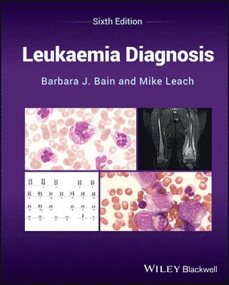
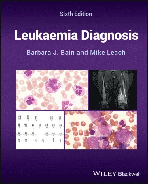
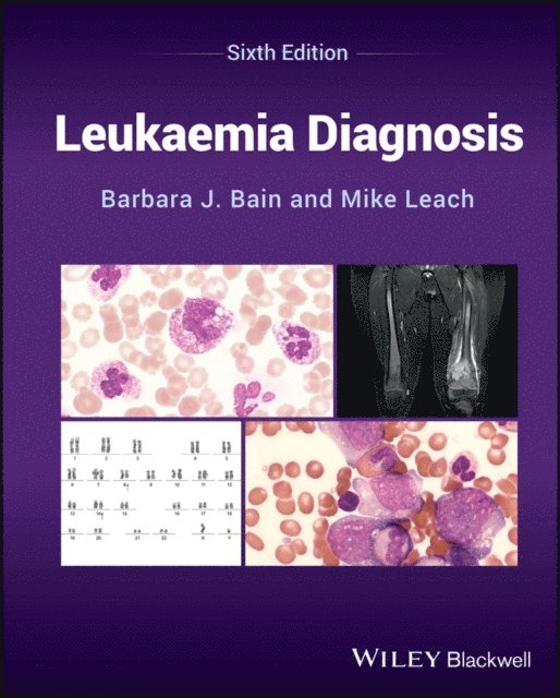
Leukaemia Diagnosis is a practical reference on the principles of leukaemia diagnosis and classification that illustrates and explains in a user-friendly way how different laboratory techniques are used to achieve an accurate interpretation. To aid in reader comprehension, over 300 high quality full colour digital images of abnormal cells in leukaemia and lymphoma are included, supplemented by histological, cytogenetic and immunophenotyping images.
This newly revised and updated Sixth Edition includes recent developments, highlights the growing importance of molecular genetics, and incorporates the recent 5th edition of the WHO guidelines and the International Consensus Classification for leukaemia diagnosis and classification throughout the text. Information on cytogenetic and molecular genetic abnormalities in leukaemia is also included, along with characteristic immunophenotypic characteristics of different categories of leukaemia.
Written by world-renowned authors in the field, Barbara Bain and Mike Leach, Leukaemia Diagnosis covers sample topics such as:
The nature of leukaemia, cytology, cytochemistry, and the morphological classification of acute leukaemia, with an index of commonly used abbreviations Immunophenotyping and cytogenetic/molecular genetic analysis, and integration of morphological, immunophenotypic and genetic information with the WHO classifications Acute lymphoblastic leukaemia, acute leukaemia of ambiguous lineage, and the myelodysplastic syndromes and myelodysplastic/myeloproliferative neoplasms Chronic myeloid leukaemias, lymphoid leukaemias of mature B, T, and natural killer cells, and leukaemia diagnosis in resource-poor countriesThe Sixth Edition of Leukaemia Diagnosis is a highly valuable resource for trainee haematologists and laboratory scientists in haematology and related disciplines. The text also serves as a useful reference and teaching aid for those who already have expertise in this field.
2 509 kr
Läs direkt efter köp
Authoritative reference on classifying and diagnosing leukaemia, with practical guidance on using various laboratory techniques included
Leukaemia Diagnosis is a practical reference on the principles of leukaemia diagnosis and classification that illustrates and explains in a user-friendly way how different laboratory techniques are used to achieve an accurate interpretation. To aid in reader comprehension, over 300 high quality full colour digital images of abnormal cells in leukaemia and lymphoma are included, supplemented by histological, cytogenetic and immunophenotyping images.
This newly revised and updated Sixth Edition includes recent developments, highlights the growing importance of molecular genetics, and incorporates the recent 5th edition of the WHO guidelines and the International Consensus Classification for leukaemia diagnosis and classification throughout the text. Information on cytogenetic and molecular genetic abnormalities in leukaemia is also included, along with characteristic immunophenotypic characteristics of different categories of leukaemia.
Written by world-renowned authors in the field, Barbara Bain and Mike Leach, Leukaemia Diagnosis covers sample topics such as:
The nature of leukaemia, cytology, cytochemistry, and the morphological classification of acute leukaemia, with an index of commonly used abbreviations Immunophenotyping and cytogenetic/molecular genetic analysis, and integration of morphological, immunophenotypic and genetic information with the WHO classifications Acute lymphoblastic leukaemia, acute leukaemia of ambiguous lineage, and the myelodysplastic syndromes and myelodysplastic/myeloproliferative neoplasms Chronic myeloid leukaemias, lymphoid leukaemias of mature B, T, and natural killer cells, and leukaemia diagnosis in resource-poor countriesThe Sixth Edition of Leukaemia Diagnosis is a highly valuable resource for trainee haematologists and laboratory scientists in haematology and related disciplines. The text also serves as a useful reference and teaching aid for those who already have expertise in this field.
1 662 kr
Skickas inom 5-8 vardagar
1 922 kr
Läs direkt efter köp
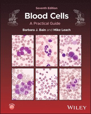
A comprehensive guide to blood cell morphology and interpretation, fully updated throughout
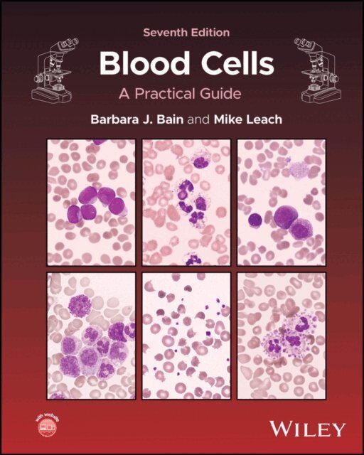
Understanding blood cell morphology and interpreting blood counts accurately is essential for both haematologists and biomedical scientists. This meticulously updated and comprehensive seventh edition of Blood Cells: A Practical Guide combines expert guidance with high-quality imaging. This edition introduces new clinical images to enhance the contextual understanding of haematological findings, alongside the latest WHO and International Consensus classifications of haematological neoplasms.
Blood Cells: A Practical Guide bridges the gap between laboratory science and clinical application, covering essential techniques such as specimen collection, blood film preparation and staining. The authors also discuss the principles of manual and automated blood counts, as well as abnormalities in blood counts and films within a clinical context. With up-to-date advice on additional tests to aid diagnosis, this new edition reinforces the importance of microscopy whilst addressing the evolving demands of laboratory and clinical haematology.
Designed as both a reference for practising haematologists and a bench manual for trainees, Blood Cells: A Practical Guide:
Features new high-quality clinical and morphological images to enhance blood cell identification and interpretation Highlights potential pitfalls and spurious results in haematological analysis to improve diagnostic accuracy Offers a self-assessment component with multiple choice questions (MCQs) and extended matching questions (EMQs) available online Is written by leading experts with decades of experience in diagnostic haematology and blood cell morphologyBlood Cells: A Practical Guide remains an indispensable resource for those dedicated to the study and practice of haematology, such as haematologists, clinical scientists, and biomedical scientists at all levels. It also serves as a valuable reference for diagnostic haematology courses and laboratory training programmes in medical and biomedical science degrees.
1 855 kr
Läs direkt efter köp
A comprehensive guide to blood cell morphology and interpretation, fully updated throughout
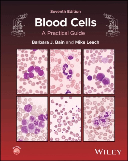
Understanding blood cell morphology and interpreting blood counts accurately is essential for both haematologists and biomedical scientists. This meticulously updated and comprehensive seventh edition of Blood Cells: A Practical Guide combines expert guidance with high-quality imaging. This edition introduces new clinical images to enhance the contextual understanding of haematological findings, alongside the latest WHO and International Consensus classifications of haematological neoplasms.
Blood Cells: A Practical Guide bridges the gap between laboratory science and clinical application, covering essential techniques such as specimen collection, blood film preparation and staining. The authors also discuss the principles of manual and automated blood counts, as well as abnormalities in blood counts and films within a clinical context. With up-to-date advice on additional tests to aid diagnosis, this new edition reinforces the importance of microscopy whilst addressing the evolving demands of laboratory and clinical haematology.
Designed as both a reference for practising haematologists and a bench manual for trainees, Blood Cells: A Practical Guide:
Features new high-quality clinical and morphological images to enhance blood cell identification and interpretation Highlights potential pitfalls and spurious results in haematological analysis to improve diagnostic accuracy Offers a self-assessment component with multiple choice questions (MCQs) and extended matching questions (EMQs) available online Is written by leading experts with decades of experience in diagnostic haematology and blood cell morphologyBlood Cells: A Practical Guide remains an indispensable resource for those dedicated to the study and practice of haematology, such as haematologists, clinical scientists, and biomedical scientists at all levels. It also serves as a valuable reference for diagnostic haematology courses and laboratory training programmes in medical and biomedical science degrees.
244 kr
Skickas inom 3-6 vardagar
146 kr
Läs direkt efter köp
189 kr
Skickas inom 5-8 vardagar